 Movie
Movie Controller


+ Open data

- Basic information
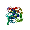
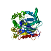
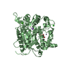
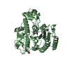
| Entry | Database: PDB / ID: 2z5g | ||||||
|---|---|---|---|---|---|---|---|
| Title | Crystal structure of T1 lipase F16L mutant | ||||||
 Components Components | Thermostable lipase | ||||||
 Keywords Keywords | HYDROLASE / lipase / cation-pi interaction | ||||||
| Function / homology |  Function and homology information Function and homology informationtriacylglycerol lipase / triacylglycerol lipase activity / lipid catabolic process / extracellular region / metal ion binding Similarity search - Function | ||||||
| Biological species |  Geobacillus zalihae (bacteria) Geobacillus zalihae (bacteria) | ||||||
| Method |  X-RAY DIFFRACTION / MOLECULAR REPLACEMENT / Resolution: 1.8 Å X-RAY DIFFRACTION / MOLECULAR REPLACEMENT / Resolution: 1.8 Å | ||||||
 Authors Authors | Matsumura, H. / Yamamoto, T. / Inoue, T. / Kai, Y. | ||||||
 Citation Citation | Journal: Proteins / Year: 2007 Title: Novel cation-pi interaction revealed by crystal structure of thermoalkalophilic lipase Authors: Matsumura, H. / Yamamoto, T. / Leow, T.C. / Mori, T. / Salleh, A.B. / Basri, M. / Inoue, T. / Kai, Y. / Rahman, R.N.Z.R.A. | ||||||
| History |
|
- Structure visualization
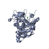
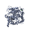
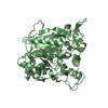
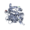
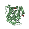
| Structure viewer | Molecule: MolmilJmol/JSmol |
|---|
- Downloads & links
-Download
| PDBx/mmCIF format | 2z5g.cif.gz | 175.7 KB | Display | PDBx/mmCIF format |
|---|---|---|---|---|
| PDB format | pdb2z5g.ent.gz | 138.4 KB | Display | PDB format |
| PDBx/mmJSON format | 2z5g.json.gz | Tree view | PDBx/mmJSON format | |
| Others |  Other downloads Other downloads |
-Validation report
| Arichive directory | https://data.pdbj.org/pub/pdb/validation_reports/z5/2z5gftp://data.pdbj.org/pub/pdb/validation_reports/z5/2z5g | HTTPS FTP |
|---|
-Related structure data
| Related structure data |  2dsnSC S: Starting model for refinement C: citing same article ( |
|---|---|
| Similar structure data |

-Links
 PDBj
PDBj- Assembly
| Deposited unit | 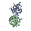
| |||||||||
|---|---|---|---|---|---|---|---|---|---|---|
| 1 | 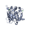
| |||||||||
| 2 | 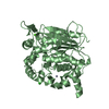
| |||||||||
| Unit cell |
| |||||||||
| Components on special symmetry positions |
|
-Components
| #1: Protein | Mass: 43155.051 Da / Num. of mol.: 2 / Fragment: UNP residues 30-416 / Mutation: F16L Source method: isolated from a genetically manipulated source Source: (gene. exp.) Geobacillus zalihae (bacteria) / Plasmid: pGEX/T1S / Production host: #2: Chemical |   Mass: 65.409 Da / Num. of mol.: 2 / Source method: obtained synthetically / Formula: Zn Mass: 65.409 Da / Num. of mol.: 2 / Source method: obtained synthetically / Formula: Zn#3: Chemical |   Mass: 40.078 Da / Num. of mol.: 2 / Source method: obtained synthetically / Formula: Ca Mass: 40.078 Da / Num. of mol.: 2 / Source method: obtained synthetically / Formula: Ca#4: Chemical |   Mass: 35.453 Da / Num. of mol.: 2 / Source method: obtained synthetically / Formula: Cl Mass: 35.453 Da / Num. of mol.: 2 / Source method: obtained synthetically / Formula: Cl#5: Water | ChemComp-HOH / |  Mass: 18.015 Da / Num. of mol.: 686 / Source method: isolated from a natural source / Formula: H2O Mass: 18.015 Da / Num. of mol.: 686 / Source method: isolated from a natural source / Formula: H2O |
|---|
-Experimental details
-Experiment
| Experiment | Method: X-RAY DIFFRACTION / Number of used crystals: 1 |
|---|
- Sample preparation
| Crystal | Density Matthews: 2.73 Å3/Da / Density % sol: 54.97 % |
|---|---|
| Crystal grow | Temperature: 293 K / Method: vapor diffusion, hanging drop / pH: 6.6 Details: 0.5M NaCl, 0.1M KH2PO4, 0.1M NaH2PO4, 0.1M MES buffer, pH 6.6, VAPOR DIFFUSION, HANGING DROP, temperature 293K |
-Data collection
| Diffraction | Mean temperature: 100 K |
|---|---|
| Diffraction source | Source: ROTATING ANODE / Type: RIGAKU ULTRAX 18 / Wavelength: 1.54 Å |
| Detector | Type: RIGAKU RAXIS IV++ / Detector: IMAGE PLATE |
| Radiation | Monochromator: YALE MIRRORS / Protocol: SINGLE WAVELENGTH / Monochromatic (M) / Laue (L): M / Scattering type: x-ray |
| Radiation wavelength | Wavelength: 1.54 Å / Relative weight: 1 |
| Reflection | Resolution: 1.8→32.3 Å / Num. obs: 83050 / % possible obs: 96.5 % / Observed criterion σ(I): -3 / Redundancy: 2.3 % / Biso Wilson estimate: 16.1 Å2 / Rmerge(I) obs: 0.06 / Net I/σ(I): 14.3 |
| Reflection shell | Resolution: 1.8→1.86 Å / Redundancy: 2.2 % / Rmerge(I) obs: 0.344 / Num. unique all: 8179 / % possible all: 95.7 |
- Processing
| Software |
| ||||||||||||||||||||||||||||||||||||||||||||||||||||||||||||||||||||||||||||||||
|---|---|---|---|---|---|---|---|---|---|---|---|---|---|---|---|---|---|---|---|---|---|---|---|---|---|---|---|---|---|---|---|---|---|---|---|---|---|---|---|---|---|---|---|---|---|---|---|---|---|---|---|---|---|---|---|---|---|---|---|---|---|---|---|---|---|---|---|---|---|---|---|---|---|---|---|---|---|---|---|---|---|
| Refinement | Method to determine structure: MOLECULAR REPLACEMENT Starting model: PDB ENTRY 2DSN Resolution: 1.8→32.25 Å / Rfactor Rfree error: 0.003 / Data cutoff high absF: 393813.08 / Data cutoff low absF: 0 / Isotropic thermal model: RESTRAINED / Cross valid method: THROUGHOUT / σ(F): 0
| ||||||||||||||||||||||||||||||||||||||||||||||||||||||||||||||||||||||||||||||||
| Solvent computation | Solvent model: FLAT MODEL / Bsol: 52.1305 Å2 / ksol: 0.374752 e/Å3 | ||||||||||||||||||||||||||||||||||||||||||||||||||||||||||||||||||||||||||||||||
| Displacement parameters | Biso mean: 21.6 Å2
| ||||||||||||||||||||||||||||||||||||||||||||||||||||||||||||||||||||||||||||||||
| Refine analyze |
| ||||||||||||||||||||||||||||||||||||||||||||||||||||||||||||||||||||||||||||||||
| Refinement step | Cycle: LAST / Resolution: 1.8→32.25 Å
| ||||||||||||||||||||||||||||||||||||||||||||||||||||||||||||||||||||||||||||||||
| Refine LS restraints |
| ||||||||||||||||||||||||||||||||||||||||||||||||||||||||||||||||||||||||||||||||
| LS refinement shell | Resolution: 1.8→1.91 Å / Rfactor Rfree error: 0.012 / Total num. of bins used: 6
| ||||||||||||||||||||||||||||||||||||||||||||||||||||||||||||||||||||||||||||||||
| Xplor file |
|
