| 登録情報 | データベース: PDB / ID: 2z4j
|
|---|
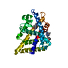
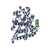
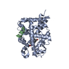
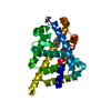
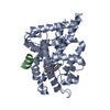
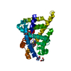
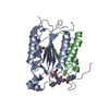
| タイトル | Crystal structure of AR LBD with SHP peptide NR Box 2 |
|---|
 要素 要素 | - Androgen receptor
- Nuclear receptor 0B2
|
|---|
 キーワード キーワード | TRANSCRIPTION / Androgen receptor / ligand binding domain / SHP / Co-repressor |
|---|
| 機能・相同性 |  機能・相同性情報 機能・相同性情報
male somatic sex determination / prostate induction / lateral sprouting involved in mammary gland duct morphogenesis / regulation of developmental growth / male genitalia morphogenesis / POU domain binding / positive regulation of integrin biosynthetic process / tertiary branching involved in mammary gland duct morphogenesis / cellular response to testosterone stimulus / animal organ formation ...male somatic sex determination / prostate induction / lateral sprouting involved in mammary gland duct morphogenesis / regulation of developmental growth / male genitalia morphogenesis / POU domain binding / positive regulation of integrin biosynthetic process / tertiary branching involved in mammary gland duct morphogenesis / cellular response to testosterone stimulus / animal organ formation / androgen binding / regulation of systemic arterial blood pressure / Leydig cell differentiation / epithelial cell differentiation involved in prostate gland development / positive regulation of epithelial cell proliferation involved in prostate gland development / prostate gland epithelium morphogenesis / prostate gland growth / epithelial cell morphogenesis / membraneless organelle assembly / RNA polymerase II general transcription initiation factor binding / positive regulation of insulin-like growth factor receptor signaling pathway / peroxisome proliferator activated receptor binding / nuclear thyroid hormone receptor binding / androgen receptor signaling pathway / positive regulation of transcription by RNA polymerase III / morphogenesis of an epithelial fold / cellular response to steroid hormone stimulus / positive regulation of intracellular estrogen receptor signaling pathway / seminiferous tubule development / RUNX2 regulates osteoblast differentiation / nuclear steroid receptor activity / mammary gland alveolus development / cellular response to estrogen stimulus / estrogen response element binding / animal organ regeneration / : / single fertilization / nuclear retinoid X receptor binding / RNA polymerase II core promoter sequence-specific DNA binding / response to glucose / cholesterol metabolic process / transcription regulator inhibitor activity / regulation of protein localization to plasma membrane / estrogen receptor signaling pathway / intracellular receptor signaling pathway / steroid binding / Notch signaling pathway / insulin-like growth factor receptor signaling pathway / epithelial cell proliferation / HSP90 chaperone cycle for steroid hormone receptors (SHR) in the presence of ligand / nuclear receptor binding / negative regulation of extrinsic apoptotic signaling pathway / SUMOylation of intracellular receptors / RNA polymerase II transcription regulatory region sequence-specific DNA binding / circadian regulation of gene expression / positive regulation of cell differentiation / molecular condensate scaffold activity / circadian rhythm / Activated PKN1 stimulates transcription of AR (androgen receptor) regulated genes KLK2 and KLK3 / Nuclear Receptor transcription pathway / positive regulation of miRNA transcription / beta-catenin binding / male gonad development / nuclear receptor activity / multicellular organism growth / transcription coactivator binding / negative regulation of epithelial cell proliferation / positive regulation of insulin secretion / transcription corepressor activity / MAPK cascade / cell-cell signaling / ATPase binding / DNA-binding transcription activator activity, RNA polymerase II-specific / spermatogenesis / transcription by RNA polymerase II / in utero embryonic development / molecular adaptor activity / RNA polymerase II-specific DNA-binding transcription factor binding / response to ethanol / DNA-binding transcription factor activity, RNA polymerase II-specific / positive regulation of MAPK cascade / nuclear speck / transcription cis-regulatory region binding / Ub-specific processing proteases / RNA polymerase II cis-regulatory region sequence-specific DNA binding / DNA-binding transcription factor activity / signaling receptor binding / negative regulation of cell population proliferation / protein domain specific binding / negative regulation of gene expression / negative regulation of DNA-templated transcription / positive regulation of cell population proliferation / chromatin binding / positive regulation of gene expression / positive regulation of DNA-templated transcription / chromatin / protein-containing complex binding / enzyme binding / negative regulation of transcription by RNA polymerase II / signal transduction類似検索 - 分子機能 Nuclear receptor subfamily 0 group B member 1/2 / Androgen receptor / Androgen receptor / : / Retinoid X Receptor / Retinoid X Receptor / Nuclear hormone receptor / Nuclear hormones receptors DNA-binding region signature. / Zinc finger, nuclear hormone receptor-type / Double treble clef zinc finger, C4 type ...Nuclear receptor subfamily 0 group B member 1/2 / Androgen receptor / Androgen receptor / : / Retinoid X Receptor / Retinoid X Receptor / Nuclear hormone receptor / Nuclear hormones receptors DNA-binding region signature. / Zinc finger, nuclear hormone receptor-type / Double treble clef zinc finger, C4 type / Nuclear hormone receptors DNA-binding domain profile. / c4 zinc finger in nuclear hormone receptors / Nuclear hormone receptor, ligand-binding domain / Nuclear hormone receptor-like domain superfamily / Ligand-binding domain of nuclear hormone receptor / Nuclear receptor (NR) ligand-binding (LBD) domain profile. / Ligand binding domain of hormone receptors / Zinc finger, NHR/GATA-type / Orthogonal Bundle / Mainly Alpha類似検索 - ドメイン・相同性 5-ALPHA-DIHYDROTESTOSTERONE / Androgen receptor / Nuclear receptor subfamily 0 group B member 2類似検索 - 構成要素 |
|---|
| 生物種 |  Homo sapiens (ヒト) Homo sapiens (ヒト) |
|---|
| 手法 |  X線回折 / シンクロトロン / 分子置換 / 解像度: 2.6 Å X線回折 / シンクロトロン / 分子置換 / 解像度: 2.6 Å |
|---|
 データ登録者 データ登録者 | Jouravel, N. / Fletterick, R.J. |
|---|
 引用 引用 | ジャーナル: Acta Crystallogr.,Sect.D / 年: 2007
タイトル: Interaction between the androgen receptor and a segment of its corepressor SHP
著者: Jouravel, N. / Sablin, E. / Arnold, L.A. / Guy, R.K. / Fletterick, R.J. |
|---|
| 履歴 | | 登録 | 2007年6月19日 | 登録サイト: PDBJ / 処理サイト: PDBJ |
|---|
| 改定 1.0 | 2007年12月4日 | Provider: repository / タイプ: Initial release |
|---|
| 改定 1.1 | 2011年7月13日 | Group: Version format compliance |
|---|
| 改定 1.2 | 2023年11月1日 | Group: Data collection / Database references ...Data collection / Database references / Derived calculations / Refinement description
カテゴリ: chem_comp_atom / chem_comp_bond ...chem_comp_atom / chem_comp_bond / database_2 / pdbx_initial_refinement_model / struct_site
Item: _database_2.pdbx_DOI / _database_2.pdbx_database_accession ..._database_2.pdbx_DOI / _database_2.pdbx_database_accession / _struct_site.pdbx_auth_asym_id / _struct_site.pdbx_auth_comp_id / _struct_site.pdbx_auth_seq_id |
|---|
|
|---|
 ムービー
ムービー コントローラー


 データを開く
 基本情報
基本情報 構造の表示
構造の表示 ダウンロードとリンク
 その他のダウンロード


 PDBj
PDBj


 集合体
集合体

 分子量: 290.440 Da / 分子数: 1 / 由来タイプ: 合成 / 式: C19H30O2
 分子量: 18.015 Da / 分子数: 6 / 由来タイプ: 天然 / 式: H2O
 試料調製
試料調製 / ビームライン: 8.3.1 / 波長: 1.11589 Å
 解析
解析