 Movie
Movie Controller

[English] 日本語
 Yorodumi
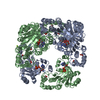
- PDB-2yw7: Crystal structure of C-terminal deletion mutant of Mycobacterium ... -
+ Open data

- Basic information
| Entry | Database: PDB / ID: 2yw7 | ||||||
|---|---|---|---|---|---|---|---|
| Title | Crystal structure of C-terminal deletion mutant of Mycobacterium smegmatis Dps | ||||||
 Components Components | Starvation-induced DNA protecting protein | ||||||
 Keywords Keywords | DNA BINDING PROTEIN / DNA-BINDING PROTEIN / Quarternary assebmly | ||||||
| Function / homology |  Function and homology information Function and homology informationOxidoreductases; Oxidizing metal ions / oxidoreductase activity, acting on metal ions / nucleoid / ferric iron binding / intracellular iron ion homeostasis / DNA binding Similarity search - Function | ||||||
| Biological species |  Mycobacterium smegmatis (bacteria) Mycobacterium smegmatis (bacteria) | ||||||
| Method |  X-RAY DIFFRACTION / MOLECULAR REPLACEMENT / Resolution: 3.3 Å X-RAY DIFFRACTION / MOLECULAR REPLACEMENT / Resolution: 3.3 Å | ||||||
 Authors Authors | Roy, S. / Saraswathi, R. / Gupta, S. / Sekar, K. / Chatterji, D. / Vijayan, M. | ||||||
 Citation Citation | Journal: J.Mol.Biol. / Year: 2007 Title: Role of N and C-terminal Tails in DNA Binding and Assembly in Dps: Structural Studies of Mycobacterium smegmatis Dps Deletion Mutants Authors: Roy, S. / Saraswathi, R. / Gupta, S. / Sekar, K. / Chatterji, D. / Vijayan, M. | ||||||
| History |
|
- Structure visualization
| Structure viewer | Molecule: MolmilJmol/JSmol |
|---|
- Downloads & links
-Download
| PDBx/mmCIF format | 2yw7.cif.gz | 285.6 KB | Display | PDBx/mmCIF format |
|---|---|---|---|---|
| PDB format | pdb2yw7.ent.gz | 233.6 KB | Display | PDB format |
| PDBx/mmJSON format | 2yw7.json.gz | Tree view | PDBx/mmJSON format | |
| Others |  Other downloads Other downloads |
-Validation report
| Arichive directory | https://data.pdbj.org/pub/pdb/validation_reports/yw/2yw7ftp://data.pdbj.org/pub/pdb/validation_reports/yw/2yw7 | HTTPS FTP |
|---|
-Related structure data
| Related structure data | 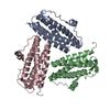 2yw6C 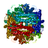 1veiS S: Starting model for refinement C: citing same article ( |
|---|---|
| Similar structure data |


-Links
 PDBj
PDBj


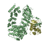
- Assembly
| Deposited unit | 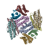
| ||||||||
|---|---|---|---|---|---|---|---|---|---|
| 1 |
| ||||||||
| Unit cell |
|
-Components
| #1: Protein | Mass: 20298.809 Da / Num. of mol.: 10 Source method: isolated from a genetically manipulated source Source: (gene. exp.) Mycobacterium smegmatis (bacteria) / Plasmid: pRSET-msdpsdeltaC26 / Species (production host): Escherichia coli / Production host: |
|---|
-Experimental details
-Experiment
| Experiment | Method: X-RAY DIFFRACTION / Number of used crystals: 1 |
|---|
- Sample preparation
| Crystal | Density Matthews: 2.15 Å3/Da / Density % sol: 42.72 % |
|---|---|
| Crystal grow | Temperature: 293 K / Method: vapor diffusion, hanging drop / pH: 8.5 Details: 30% PEG1140, 100mM CaCl2, 0.1M Tris-HCl pH8.5, VAPOR DIFFUSION, HANGING DROP, temperature 293K |
-Data collection
| Diffraction | Mean temperature: 293 K |
|---|---|
| Diffraction source | Source: ROTATING ANODE / Type: RIGAKU RU200 / Wavelength: 1.5418 Å |
| Detector | Type: MAR scanner 300 mm plate / Detector: IMAGE PLATE / Date: Jan 20, 2006 |
| Radiation | Monochromator: OSMIC MIRROR / Protocol: SINGLE WAVELENGTH / Monochromatic (M) / Laue (L): M / Scattering type: x-ray |
| Radiation wavelength | Wavelength: 1.5418 Å / Relative weight: 1 |
| Reflection | Resolution: 3.3→30 Å / Num. obs: 24653 / % possible obs: 100 % / Redundancy: 4.1 % / Rmerge(I) obs: 0.0108 / Net I/σ(I): 11.7 |
| Reflection shell | Resolution: 3.3→3.382 Å / Rmerge(I) obs: 0.0331 / Mean I/σ(I) obs: 3.3 / Num. unique all: 2467 / % possible all: 100 |
- Processing
| Software |
| |||||||||||||||||||||||||||||||||||||||||||||||||||||||||||||||||||||||||||||||||||||||||||||||
|---|---|---|---|---|---|---|---|---|---|---|---|---|---|---|---|---|---|---|---|---|---|---|---|---|---|---|---|---|---|---|---|---|---|---|---|---|---|---|---|---|---|---|---|---|---|---|---|---|---|---|---|---|---|---|---|---|---|---|---|---|---|---|---|---|---|---|---|---|---|---|---|---|---|---|---|---|---|---|---|---|---|---|---|---|---|---|---|---|---|---|---|---|---|---|---|---|
| Refinement | Method to determine structure: MOLECULAR REPLACEMENT Starting model: 1VEI Resolution: 3.3→28.54 Å / Cor.coef. Fo:Fc: 0.903 / Cor.coef. Fo:Fc free: 0.874 / SU B: 23.764 / SU ML: 0.399 / Cross valid method: THROUGHOUT / ESU R Free: 0.598 / Stereochemistry target values: MAXIMUM LIKELIHOOD / Details: HYDROGENS HAVE BEEN ADDED IN THE RIDING POSITIONS
| |||||||||||||||||||||||||||||||||||||||||||||||||||||||||||||||||||||||||||||||||||||||||||||||
| Solvent computation | Ion probe radii: 0.8 Å / Shrinkage radii: 0.8 Å / VDW probe radii: 1.2 Å / Solvent model: BABINET MODEL WITH MASK | |||||||||||||||||||||||||||||||||||||||||||||||||||||||||||||||||||||||||||||||||||||||||||||||
| Displacement parameters | Biso mean: 43.228 Å2
| |||||||||||||||||||||||||||||||||||||||||||||||||||||||||||||||||||||||||||||||||||||||||||||||
| Refine analyze |
| |||||||||||||||||||||||||||||||||||||||||||||||||||||||||||||||||||||||||||||||||||||||||||||||
| Refinement step | Cycle: LAST / Resolution: 3.3→28.54 Å
| |||||||||||||||||||||||||||||||||||||||||||||||||||||||||||||||||||||||||||||||||||||||||||||||
| Refine LS restraints |
| |||||||||||||||||||||||||||||||||||||||||||||||||||||||||||||||||||||||||||||||||||||||||||||||
| LS refinement shell | Resolution: 3.297→3.382 Å / Total num. of bins used: 20
|
