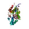
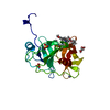
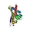
 Movie
Movie Controller


+ Open data

- Basic information
| Entry | Database: PDB / ID: 2yoq | ||||||
|---|---|---|---|---|---|---|---|
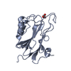
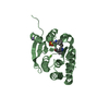
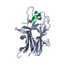
| Title | Structure of FAM3B PANDER E30 construct | ||||||
 Components Components | PROTEIN FAM3B | ||||||
 Keywords Keywords | APOPTOSIS / DIABETES / ILEI / EMT | ||||||
| Function / homology |  Function and homology information Function and homology informationinsulin secretion / nuclear envelope lumen / cytokine activity / carbohydrate binding / apoptotic process / signal transduction / : / extracellular region Similarity search - Function | ||||||
| Biological species |  | ||||||
| Method |  X-RAY DIFFRACTION / SYNCHROTRON / MOLECULAR REPLACEMENT / Resolution: 2.35 Å X-RAY DIFFRACTION / SYNCHROTRON / MOLECULAR REPLACEMENT / Resolution: 2.35 Å | ||||||
 Authors Authors | Johansson, P. / Bernstrom, J. / Gorman, T. / Oster, L. / Backstrom, S. / Schweikart, F. / Xu, B. / Xue, Y. / Holmberg Schiavone, L. | ||||||
 Citation Citation | Journal: Structure / Year: 2013 Title: Fam3B Pander and Fam3C Ilei Represent a Distinct Class of Signaling Molecules with a Non-Cytokine-Like Fold. Authors: Johansson, P. / Bernstrom, J. / Gorman, T. / Oster, L. / Backstrom, S. / Schweikart, F. / Xu, B. / Xue, Y. / Schiavone, L.H. | ||||||
| History |
|
- Structure visualization
| Structure viewer | Molecule: MolmilJmol/JSmol |
|---|
- Downloads & links
-Download
| PDBx/mmCIF format | 2yoq.cif.gz | 127.2 KB | Display | PDBx/mmCIF format |
|---|---|---|---|---|
| PDB format | pdb2yoq.ent.gz | 99 KB | Display | PDB format |
| PDBx/mmJSON format | 2yoq.json.gz | Tree view | PDBx/mmJSON format | |
| Others |  Other downloads Other downloads |
-Validation report
| Arichive directory | https://data.pdbj.org/pub/pdb/validation_reports/yo/2yoqftp://data.pdbj.org/pub/pdb/validation_reports/yo/2yoq | HTTPS FTP |
|---|
-Related structure data


-Links
 PDBj
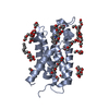
PDBj- Assembly
| Deposited unit | 
| ||||||||||||
|---|---|---|---|---|---|---|---|---|---|---|---|---|---|
| 1 | 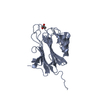
| ||||||||||||
| 2 | 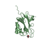
| ||||||||||||
| 3 | 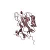
| ||||||||||||
| Unit cell |
| ||||||||||||
| Noncrystallographic symmetry (NCS) | NCS oper:
|
-Components
| #1: Protein | Mass: 23974.236 Da / Num. of mol.: 3 Source method: isolated from a genetically manipulated source Source: (gene. exp.)  HOMO SAPIENS (human) / References: UniProt: Q9D309 HOMO SAPIENS (human) / References: UniProt: Q9D309#2: Chemical |   Mass: 92.094 Da / Num. of mol.: 3 / Source method: obtained synthetically / Formula: C3H8O3 Mass: 92.094 Da / Num. of mol.: 3 / Source method: obtained synthetically / Formula: C3H8O3#3: Water | ChemComp-HOH / |  Mass: 18.015 Da / Num. of mol.: 412 / Source method: isolated from a natural source / Formula: H2O Mass: 18.015 Da / Num. of mol.: 412 / Source method: isolated from a natural source / Formula: H2OHas protein modification | Y | |
|---|
-Experimental details
-Experiment
| Experiment | Method: X-RAY DIFFRACTION / Number of used crystals: 1 |
|---|
- Sample preparation
| Crystal | Density Matthews: 3.2 Å3/Da / Density % sol: 61.13 % / Description: NONE |
|---|---|
| Crystal grow | pH: 7 / Details: pH 7 |
-Data collection
| Diffraction | Mean temperature: 100 K |
|---|---|
| Diffraction source | Source: SYNCHROTRON / Site: ESRF  / Beamline: ID23-1 / Wavelength: 1.072 / Beamline: ID23-1 / Wavelength: 1.072 |
| Detector | Type: ADSC CCD / Detector: CCD / Date: Nov 11, 2011 |
| Radiation | Protocol: SINGLE WAVELENGTH / Monochromatic (M) / Laue (L): M / Scattering type: x-ray |
| Radiation wavelength | Wavelength: 1.072 Å / Relative weight: 1 |
| Reflection | Resolution: 2.35→34.47 Å / Num. obs: 33513 / % possible obs: 99.9 % / Observed criterion σ(I): 1 / Redundancy: 4.3 % / Biso Wilson estimate: 42.49 Å2 / Rmerge(I) obs: 0.13 / Net I/σ(I): 11.2 |
| Reflection shell | Resolution: 2.35→2.43 Å / Redundancy: 4.1 % / Rmerge(I) obs: 0.79 / Mean I/σ(I) obs: 2.6 / % possible all: 99.7 |
- Processing
| Software |
| ||||||||||||||||||||||||||||||||||||||||||||||||||||||||||||||||||||||||||||||||||||||||||||||||||||||||||||||||||
|---|---|---|---|---|---|---|---|---|---|---|---|---|---|---|---|---|---|---|---|---|---|---|---|---|---|---|---|---|---|---|---|---|---|---|---|---|---|---|---|---|---|---|---|---|---|---|---|---|---|---|---|---|---|---|---|---|---|---|---|---|---|---|---|---|---|---|---|---|---|---|---|---|---|---|---|---|---|---|---|---|---|---|---|---|---|---|---|---|---|---|---|---|---|---|---|---|---|---|---|---|---|---|---|---|---|---|---|---|---|---|---|---|---|---|---|
| Refinement | Method to determine structure: MOLECULAR REPLACEMENT / Resolution: 2.35→21.64 Å / Cor.coef. Fo:Fc: 0.9457 / Cor.coef. Fo:Fc free: 0.9344 / SU R Cruickshank DPI: 0.211 / Cross valid method: THROUGHOUT / σ(F): 0 / SU R Blow DPI: 0.215 / SU Rfree Blow DPI: 0.167 / SU Rfree Cruickshank DPI: 0.167 Details: IDEAL-DIST CONTACT TERM CONTACT SETUP. ALL ATOMS HAVE CCP4 ATOM TYPE FROM LIBRARY
| ||||||||||||||||||||||||||||||||||||||||||||||||||||||||||||||||||||||||||||||||||||||||||||||||||||||||||||||||||
| Displacement parameters | Biso mean: 34.55 Å2
| ||||||||||||||||||||||||||||||||||||||||||||||||||||||||||||||||||||||||||||||||||||||||||||||||||||||||||||||||||
| Refine analyze | Luzzati coordinate error obs: 0.226 Å | ||||||||||||||||||||||||||||||||||||||||||||||||||||||||||||||||||||||||||||||||||||||||||||||||||||||||||||||||||
| Refinement step | Cycle: LAST / Resolution: 2.35→21.64 Å
| ||||||||||||||||||||||||||||||||||||||||||||||||||||||||||||||||||||||||||||||||||||||||||||||||||||||||||||||||||
| Refine LS restraints |
| ||||||||||||||||||||||||||||||||||||||||||||||||||||||||||||||||||||||||||||||||||||||||||||||||||||||||||||||||||
| LS refinement shell | Resolution: 2.35→2.42 Å / Total num. of bins used: 17
|
