 Movie
Movie Controller

[English] 日本語
 Yorodumi
- PDB-2yg1: APO STRUCTURE OF CELLOBIOHYDROLASE 1 (CEL7A) FROM HETEROBASIDION ... -
+ Open data

- Basic information
| Entry | Database: PDB / ID: 2yg1 | ||||||||||||
|---|---|---|---|---|---|---|---|---|---|---|---|---|---|
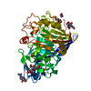
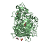
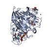
| Title | APO STRUCTURE OF CELLOBIOHYDROLASE 1 (CEL7A) FROM HETEROBASIDION ANNOSUM | ||||||||||||
 Components Components | CELLULOSE 1,4-BETA-CELLOBIOSIDASE | ||||||||||||
 Keywords Keywords | HYDROLASE / GLYCOSIDE HYDROLASE / CELLULASE / WHITE-ROT FUNGUS / BASIDIOMYCETE / FOREST PATHOGEN | ||||||||||||
| Function / homology |  Function and homology information Function and homology informationHydrolases; Glycosylases; Glycosidases, i.e. enzymes that hydrolyse O- and S-glycosyl compounds / cellulose catabolic process / hydrolase activity, hydrolyzing O-glycosyl compounds / metal ion binding Similarity search - Function | ||||||||||||
| Biological species |  HETEROBASIDION ANNOSUM (fungus) HETEROBASIDION ANNOSUM (fungus) | ||||||||||||
| Method |  X-RAY DIFFRACTION / SYNCHROTRON / MOLECULAR REPLACEMENT / Resolution: 1.9 Å X-RAY DIFFRACTION / SYNCHROTRON / MOLECULAR REPLACEMENT / Resolution: 1.9 Å | ||||||||||||
 Authors Authors | Haddad-Momeni, M. / Hansson, H. / Mikkelsen, N.E. / Wang, X. / Svedberg, J. / Sandgren, M. / Stahlberg, J. | ||||||||||||
 Citation Citation | Journal: J.Biol.Chem. / Year: 2013 Title: Structural, Biochemical, and Computational Characterization of the Glycoside Hydrolase Family 7 Cellobiohydrolase of the Tree-Killing Fungus Heterobasidion Irregulare. Authors: Momeni, M.H. / Payne, C.M. / Hansson, H. / Mikkelsen, N.E. / Svedberg, J. / Engstrom, A. / Sandgren, M. / Beckham, G.T. / Stahlberg, J. | ||||||||||||
| History |
|
- Structure visualization
| Structure viewer | Molecule: MolmilJmol/JSmol |
|---|
- Downloads & links
-Download
| PDBx/mmCIF format | 2yg1.cif.gz | 194.4 KB | Display | PDBx/mmCIF format |
|---|---|---|---|---|
| PDB format | pdb2yg1.ent.gz | 153.1 KB | Display | PDB format |
| PDBx/mmJSON format | 2yg1.json.gz | Tree view | PDBx/mmJSON format | |
| Others |  Other downloads Other downloads |
-Validation report
| Summary document | 2yg1_validation.pdf.gz | 1 MB | Display | wwPDB validaton report |
|---|---|---|---|---|
| Full document | 2yg1_full_validation.pdf.gz | 1 MB | Display | |
| Data in XML | 2yg1_validation.xml.gz | 42 KB | Display | |
| Data in CIF | 2yg1_validation.cif.gz | 64.2 KB | Display | |
| Arichive directory | https://data.pdbj.org/pub/pdb/validation_reports/yg/2yg1ftp://data.pdbj.org/pub/pdb/validation_reports/yg/2yg1 | HTTPS FTP |
-Related structure data
| Related structure data |  2xspC 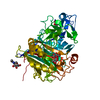 1z3vS S: Starting model for refinement C: citing same article ( |
|---|---|
| Similar structure data |


-Links
 PDBj
PDBj- Assembly
| Deposited unit | 
| ||||||||
|---|---|---|---|---|---|---|---|---|---|
| 1 | 
| ||||||||
| 2 | 
| ||||||||
| Unit cell |
|
-Components
| #1: Protein | Mass: 46991.227 Da / Num. of mol.: 2 / Source method: isolated from a natural source / Source: (natural) HETEROBASIDION ANNOSUM (fungus) / Strain: TC32-1References: UniProt: I1SB08*PLUS, Hydrolases; Glycosylases; Glycosidases, i.e. enzymes that hydrolyse O- and S-glycosyl compounds #2: Polysaccharide | Source method: isolated from a genetically manipulated source #3: Chemical |   Mass: 24.305 Da / Num. of mol.: 3 / Source method: obtained synthetically / Formula: Mg Mass: 24.305 Da / Num. of mol.: 3 / Source method: obtained synthetically / Formula: Mg#4: Water | ChemComp-HOH / |  Mass: 18.015 Da / Num. of mol.: 923 / Source method: isolated from a natural source / Formula: H2O Mass: 18.015 Da / Num. of mol.: 923 / Source method: isolated from a natural source / Formula: H2OHas protein modification | Y | |
|---|
-Experimental details
-Experiment
| Experiment | Method: X-RAY DIFFRACTION / Number of used crystals: 1 |
|---|
- Sample preparation
| Crystal | Density Matthews: 1.96 Å3/Da / Density % sol: 37 % / Description: NONE |
|---|---|
| Crystal grow | pH: 7.7 Details: PROTEIN WAS CRYSTALLIZED FROM 20 MM MGCL2, 0.1 M HEPES, PH 7.5, 22% POLYACRYLIC ACID 5100, NA SALT. |
-Data collection
| Diffraction | Mean temperature: 100 K |
|---|---|
| Diffraction source | Source: SYNCHROTRON / Site: MAX II  / Beamline: I911-2 / Wavelength: 1.04 / Beamline: I911-2 / Wavelength: 1.04 |
| Detector | Type: MARRESEARCH MAR165 / Detector: CCD / Date: Feb 15, 2010 / Details: MULTILAYER MIRROR |
| Radiation | Monochromator: BENT SI CRYSTAL / Protocol: SINGLE WAVELENGTH / Monochromatic (M) / Laue (L): M / Scattering type: x-ray |
| Radiation wavelength | Wavelength: 1.04 Å / Relative weight: 1 |
| Reflection | Resolution: 1.9→29.4 Å / Num. obs: 54342 / % possible obs: 98.1 % / Observed criterion σ(I): 3.3 / Redundancy: 4.1 % / Rmerge(I) obs: 0.06 / Net I/σ(I): 15.7 |
| Reflection shell | Resolution: 1.9→2 Å / Redundancy: 3.9 % / Rmerge(I) obs: 0.23 / Mean I/σ(I) obs: 5.5 / % possible all: 96.5 |
- Processing
| Software |
| ||||||||||||||||||||||||||||||||||||||||||||||||||||||||||||||||||||||||||||||||||||||||||||||||||||||||||||||||||||||||||||||||||||||||||||||||||||||||||||||||||||||||||||||||||||||
|---|---|---|---|---|---|---|---|---|---|---|---|---|---|---|---|---|---|---|---|---|---|---|---|---|---|---|---|---|---|---|---|---|---|---|---|---|---|---|---|---|---|---|---|---|---|---|---|---|---|---|---|---|---|---|---|---|---|---|---|---|---|---|---|---|---|---|---|---|---|---|---|---|---|---|---|---|---|---|---|---|---|---|---|---|---|---|---|---|---|---|---|---|---|---|---|---|---|---|---|---|---|---|---|---|---|---|---|---|---|---|---|---|---|---|---|---|---|---|---|---|---|---|---|---|---|---|---|---|---|---|---|---|---|---|---|---|---|---|---|---|---|---|---|---|---|---|---|---|---|---|---|---|---|---|---|---|---|---|---|---|---|---|---|---|---|---|---|---|---|---|---|---|---|---|---|---|---|---|---|---|---|---|---|
| Refinement | Method to determine structure: MOLECULAR REPLACEMENT Starting model: PDB ENTRY 1Z3V Resolution: 1.9→28.1 Å / Cor.coef. Fo:Fc: 0.948 / Cor.coef. Fo:Fc free: 0.92 / SU B: 4.495 / SU ML: 0.133 / Cross valid method: THROUGHOUT / ESU R: 0.212 / ESU R Free: 0.182 / Stereochemistry target values: MAXIMUM LIKELIHOOD Details: HYDROGENS HAVE BEEN ADDED IN THE RIDING POSITIONS. U VALUES REFINED INDIVIDUALLY. RESIDUES 45, 46, 47, 52, 53, 54, 55, 197, 198, 199, 200, 201, 202 AND 203 FROM CHAIN B, HAVE NOT BEEN MODELED.
| ||||||||||||||||||||||||||||||||||||||||||||||||||||||||||||||||||||||||||||||||||||||||||||||||||||||||||||||||||||||||||||||||||||||||||||||||||||||||||||||||||||||||||||||||||||||
| Solvent computation | Ion probe radii: 0.8 Å / Shrinkage radii: 0.8 Å / VDW probe radii: 1.4 Å / Solvent model: MASK | ||||||||||||||||||||||||||||||||||||||||||||||||||||||||||||||||||||||||||||||||||||||||||||||||||||||||||||||||||||||||||||||||||||||||||||||||||||||||||||||||||||||||||||||||||||||
| Displacement parameters | Biso mean: 20.163 Å2
| ||||||||||||||||||||||||||||||||||||||||||||||||||||||||||||||||||||||||||||||||||||||||||||||||||||||||||||||||||||||||||||||||||||||||||||||||||||||||||||||||||||||||||||||||||||||
| Refinement step | Cycle: LAST / Resolution: 1.9→28.1 Å
| ||||||||||||||||||||||||||||||||||||||||||||||||||||||||||||||||||||||||||||||||||||||||||||||||||||||||||||||||||||||||||||||||||||||||||||||||||||||||||||||||||||||||||||||||||||||
| Refine LS restraints |
|