[English] 日本語
Yorodumi
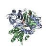
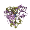
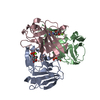
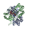
- PDB-2ybq: The x-ray structure of the SAM-dependent uroporphyrinogen III met... -

+
Open data


ID or keywords:

Loading...

-
Basic information

Entry
Database: PDB / ID: 2ybq
Title
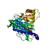
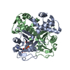
The x-ray structure of the SAM-dependent uroporphyrinogen III methyltransferase NirE from Pseudomonas aeruginosa in complex with SAH and uroporphyrinogen III
ComponentsMETHYLTRANSFERASE
KeywordsTRANSFERASE / HEME D1 BIOSYNTHESIS
Function / homology
Function and homology information


uroporphyrinogen-III C-methyltransferase / uroporphyrin-III C-methyltransferase activity / siroheme biosynthetic process / cobalamin biosynthetic process / methylation
Similarity search - Function
Uroporphyrin-III C-methyltransferase / Uroporphyrin-III C-methyltransferase signature 2. / Uroporphyrin-III C-methyltransferase signature 1. / Uroporphiryn-III C-methyltransferase, conserved site / Tetrapyrrole methylase, N-terminal domain / Tetrapyrrole methylase, C-terminal domain / Methyltransferase, Cobalt-precorrin-4 Transmethylase; Domain 2 / Tetrapyrrole methylase, subdomain 2 / Cobalt-precorrin-4 Transmethylase; domain 1 / Tetrapyrrole methylase ...Uroporphyrin-III C-methyltransferase / Uroporphyrin-III C-methyltransferase signature 2. / Uroporphyrin-III C-methyltransferase signature 1. / Uroporphiryn-III C-methyltransferase, conserved site / Tetrapyrrole methylase, N-terminal domain / Tetrapyrrole methylase, C-terminal domain / Methyltransferase, Cobalt-precorrin-4 Transmethylase; Domain 2 / Tetrapyrrole methylase, subdomain 2 / Cobalt-precorrin-4 Transmethylase; domain 1 / Tetrapyrrole methylase / Tetrapyrrole (Corrin/Porphyrin) Methylases / Tetrapyrrole methylase, subdomain 1 / Tetrapyrrole methylase superfamily / 2-Layer Sandwich / 3-Layer(aba) Sandwich / Alpha Beta
Similarity search - Domain/homology
S-ADENOSYL-L-HOMOCYSTEINE / UROPORPHYRINOGEN III / uroporphyrinogen-III C-methyltransferase
Similarity search - Component
Biological speciesPSEUDOMONAS AERUGINOSA (bacteria)
MethodX-RAY DIFFRACTION / SYNCHROTRON / MOLECULAR REPLACEMENT / Resolution: 2 Å
AuthorsStorbeck, S. / Saha, S. / Krausze, J. / Klink, B.U. / Heinz, D.W. / Layer, G.
CitationJournal: J.Biol.Chem. / Year: 2011
Title: Crystal Structure of the Heme D1 Biosynthesis Enzyme Nire in Complex with its Substrate Reveals New Insights Into the Catalytic Mechanism of S-Adenosyl-L-Methionine-Dependent Uroporphyrinogen ...Title: Crystal Structure of the Heme D1 Biosynthesis Enzyme Nire in Complex with its Substrate Reveals New Insights Into the Catalytic Mechanism of S-Adenosyl-L-Methionine-Dependent Uroporphyrinogen III Methyltransferases.
Authors: Storbeck, S. / Saha, S. / Krausze, J. / Klink, B.U. / Heinz, D.W. / Layer, G.
History
DepositionMar 9, 2011Deposition site: PDBE / Processing site: PDBE
Revision 1.0Jun 1, 2011Provider: repository / Type: Initial release
Revision 1.1Sep 28, 2011Group: Database references / Version format compliance
Revision 1.2Dec 20, 2023Group: Data collection / Database references ...Data collection / Database references / Derived calculations / Other / Refinement description
Category: chem_comp_atom / chem_comp_bond ...chem_comp_atom / chem_comp_bond / database_2 / pdbx_database_status / pdbx_initial_refinement_model / struct_site
Item: _database_2.pdbx_DOI / _database_2.pdbx_database_accession ..._database_2.pdbx_DOI / _database_2.pdbx_database_accession / _pdbx_database_status.status_code_sf / _struct_site.pdbx_auth_asym_id / _struct_site.pdbx_auth_comp_id / _struct_site.pdbx_auth_seq_id

-
Structure visualization

Structure viewerMolecule:
MolmilJmol/JSmol

Downloads & links

-
Assembly

Deposited unit
A: METHYLTRANSFERASE
hetero molecules


Theoretical massNumber of molelcules
Total (without water)32,3893
Polymers31,1681
Non-polymers1,2212
Water2,324129
1
A: METHYLTRANSFERASE
hetero molecules

A: METHYLTRANSFERASE
hetero molecules


Theoretical massNumber of molelcules
Total (without water)64,7786
Polymers62,3352
Non-polymers2,4424
Water362
TypeNameSymmetry operationNumber
identity operation1_555x,y,z1
crystal symmetry operation3_555-x,y,-z+1/21
Buried area5600 Å2
ΔGint-26.4 kcal/mol
Surface area18130 Å2
MethodPISA
Unit cell
Length a, b, c (Å)63.853, 116.702, 76.988
Angle α, β, γ (deg.)90.00, 90.00, 90.00
Int Tables number20
Space group name H-MC2221

-
Components

#1: Protein METHYLTRANSFERASE / UROPORPHYRINOGEN III METHYLTRANFERASE


Mass: 31167.736 Da / Num. of mol.: 1
Source method: isolated from a genetically manipulated source
Source: (gene. exp.) PSEUDOMONAS AERUGINOSA (bacteria) / Strain: PAO1 / Plasmid: PET32A_PRES2-4_NIRE / Production host: ESCHERICHIA COLI (E. coli) / Strain (production host): BL21(DE3)
References: UniProt: P95417, uroporphyrinogen-III C-methyltransferase
#2: Chemical ChemComp-SAH / S-ADENOSYL-L-HOMOCYSTEINE


Type: L-peptide linking / 
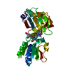
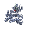
Mass: 384.411 Da / Num. of mol.: 1 / Source method: obtained synthetically / Formula: C14H20N6O5S
#3: Chemical ChemComp-UP2 / UROPORPHYRINOGEN III


Mass: 836.795 Da / Num. of mol.: 1 / Source method: obtained synthetically / Formula: C40H44N4O16
#4: Water ChemComp-HOH / water


Mass: 18.015 Da / Num. of mol.: 129 / Source method: isolated from a natural source / Formula: H2O

-
Experimental details

-
Experiment

ExperimentMethod: X-RAY DIFFRACTION / Number of used crystals: 1

-
Sample preparation

CrystalDensity Matthews: 2.44 Å3/Da / Density % sol: 46.4 % / Description: NONE
Crystal growpH: 8.5
Details: PROTEIN WAS CRYSTALLIZED FROM 1.26 M AMMONIUM SULFATE, 0.2 M LITHIUM SULFATE, 0.1 M TRIS PH 8.5

-
Data collection

DiffractionMean temperature: 100 K
Diffraction sourceSource: SYNCHROTRON / Site: BESSY / Beamline: 14.2 / Wavelength: 0.91841
DetectorType: MARRESEARCH / Detector: CCD / Date: Dec 17, 2010
RadiationMonochromator: SI(100) / Protocol: SINGLE WAVELENGTH / Monochromatic (M) / Laue (L): M / Scattering type: x-ray
Radiation wavelengthWavelength: 0.91841 Å / Relative weight: 1
ReflectionResolution: 2→35 Å / Num. obs: 18362 / % possible obs: 92.6 % / Observed criterion σ(I): 2 / Redundancy: 3.9 % / Biso Wilson estimate: 34.4 Å2 / Rmerge(I) obs: 0.08 / Net I/σ(I): 22.8
Reflection shellResolution: 2→2.05 Å / Redundancy: 4 % / Rmerge(I) obs: 0.45 / Mean I/σ(I) obs: 4 / % possible all: 99.9

-
Processing

Software
NameVersionClassification
REFMAC5.5.0109refinement
XDSdata reduction
XSCALEdata scaling
MOLREPphasing
RefinementMethod to determine structure: MOLECULAR REPLACEMENT
Starting model: PDB ENTRY 2YBO
Resolution: 2→32.13 Å / Cor.coef. Fo:Fc: 0.919 / Cor.coef. Fo:Fc free: 0.905 / SU B: 10.846 / SU ML: 0.139 / Cross valid method: THROUGHOUT / ESU R: 0.216 / ESU R Free: 0.18 / Stereochemistry target values: MAXIMUM LIKELIHOOD / Details: HYDROGENS HAVE BEEN ADDED IN THE RIDING POSITIONS.
RfactorNum. reflection% reflectionSelection details
Rfree0.25904 919 5 %RANDOM
Rwork0.22626 ---
obs0.22795 17443 100 %-
Solvent computationIon probe radii: 0.8 Å / Shrinkage radii: 0.8 Å / VDW probe radii: 1.4 Å / Solvent model: BABINET MODEL WITH MASK
Displacement parametersBiso mean: 33.641 Å2
Baniso -1Baniso -2Baniso -3
1--0.14 Å20 Å20 Å2
2--1.17 Å20 Å2
3----1.03 Å2
Refinement stepCycle: LAST / Resolution: 2→32.13 Å
ProteinNucleic acidLigandSolventTotal
Num. atoms1817 0 86 129 2032
Refine LS restraints
Refine-IDTypeDev idealDev ideal targetNumber
X-RAY DIFFRACTIONr_bond_refined_d0.0110.0221947
X-RAY DIFFRACTIONr_bond_other_d
X-RAY DIFFRACTIONr_angle_refined_deg1.3142.0272652
X-RAY DIFFRACTIONr_angle_other_deg
X-RAY DIFFRACTIONr_dihedral_angle_1_deg5.245239
X-RAY DIFFRACTIONr_dihedral_angle_2_deg36.98122.82178
X-RAY DIFFRACTIONr_dihedral_angle_3_deg14.19115298
X-RAY DIFFRACTIONr_dihedral_angle_4_deg20.2731519
X-RAY DIFFRACTIONr_chiral_restr0.0820.2301
X-RAY DIFFRACTIONr_gen_planes_refined0.0050.0211480
X-RAY DIFFRACTIONr_gen_planes_other
X-RAY DIFFRACTIONr_nbd_refined0.2010.2868
X-RAY DIFFRACTIONr_nbd_other
X-RAY DIFFRACTIONr_nbtor_refined0.3030.21277
X-RAY DIFFRACTIONr_nbtor_other
X-RAY DIFFRACTIONr_xyhbond_nbd_refined0.1290.2124
X-RAY DIFFRACTIONr_xyhbond_nbd_other
X-RAY DIFFRACTIONr_metal_ion_refined
X-RAY DIFFRACTIONr_metal_ion_other
X-RAY DIFFRACTIONr_symmetry_vdw_refined0.1790.268
X-RAY DIFFRACTIONr_symmetry_vdw_other
X-RAY DIFFRACTIONr_symmetry_hbond_refined0.1640.221
X-RAY DIFFRACTIONr_symmetry_hbond_other
X-RAY DIFFRACTIONr_symmetry_metal_ion_refined
X-RAY DIFFRACTIONr_symmetry_metal_ion_other
X-RAY DIFFRACTIONr_mcbond_it0.5631.51201
X-RAY DIFFRACTIONr_mcbond_other
X-RAY DIFFRACTIONr_mcangle_it1.02521911
X-RAY DIFFRACTIONr_mcangle_other
X-RAY DIFFRACTIONr_scbond_it1.6063746
X-RAY DIFFRACTIONr_scbond_other
X-RAY DIFFRACTIONr_scangle_it2.7214.5741
X-RAY DIFFRACTIONr_scangle_other
X-RAY DIFFRACTIONr_long_range_B_refined
X-RAY DIFFRACTIONr_long_range_B_other
X-RAY DIFFRACTIONr_rigid_bond_restr
X-RAY DIFFRACTIONr_sphericity_free
X-RAY DIFFRACTIONr_sphericity_bonded
LS refinement shellResolution: 2→2.052 Å / Total num. of bins used: 20
RfactorNum. reflection% reflection
Rfree0.34 71 -
Rwork0.222 1341 -
obs--100 %
Refinement TLS params.

Method: refined / Refine-ID: X-RAY DIFFRACTION

IDL112)L122)L132)L222)L232)L332)S11 (Å °)S12 (Å °)S13 (Å °)S21 (Å °)S22 (Å °)S23 (Å °)S31 (Å °)S32 (Å °)S33 (Å °)T112)T122)T132)T222)T232)T332)Origin x (Å)Origin y (Å)Origin z (Å)
16.19121.54860.42570.65410.33431.25340.02650.01320.5447-0.0211-0.16440.0844-0.23710.13490.13790.1287-0.03320.01790.17340.08060.245510.155235.599616.2873
28.13380.1930.89835.0714-1.6555.37440.08630.8662-0.0612-0.76440.020.5051-0.2344-0.3788-0.10630.2755-0.0579-0.03860.42640.08850.219912.094533.23615.715
31.2431.13270.64881.87220.08191.18680.05360.1815-0.1446-0.0359-0.0766-0.28030.14370.26420.0230.04550.02640.02670.12860.0210.066110.256520.962414.7548
40.549-0.44010.23131.02110.66551.41780.02280.0176-0.0026-0.1035-0.05190.05150.0682-0.09060.02920.14870.0013-0.01940.0801-0.00690.0461-5.800810.54445.2337
56.3324-4.1138-0.851213.066616.915325.89370.22941.29010.0014-1.1066-0.2630.0739-1.35470.910.03360.59090.1485-0.08350.53540.08070.4499-17.33426.70329.0634
Refinement TLS group
IDRefine-IDRefine TLS-IDAuth asym-IDAuth seq-ID
1X-RAY DIFFRACTION1A13 - 49
2X-RAY DIFFRACTION2A50 - 82
3X-RAY DIFFRACTION3A83 - 171
4X-RAY DIFFRACTION4A172 - 255
5X-RAY DIFFRACTION5A256 - 265

+
About Yorodumi

-
News

-
Feb 9, 2022. New format data for meta-information of EMDB entries

New format data for meta-information of EMDB entries

  • Version 3 of the EMDB header file is now the official format.
  • The previous official version 1.9 will be removed from the archive.

Related info.:EMDB header

External links:wwPDB to switch to version 3 of the EMDB data model

-
Aug 12, 2020. Covid-19 info

Covid-19 info

URL: https://pdbjlvh1.pdbj.org/emnavi/covid19.php

New page: Covid-19 featured information page in EM Navigator.

Related info.:Covid-19 info / Mar 5, 2020. Novel coronavirus structure data

+
Mar 5, 2020. Novel coronavirus structure data

Novel coronavirus structure data

Related info.:Yorodumi Speices / Aug 12, 2020. Covid-19 info

External links:COVID-19 featured content - PDBj / Molecule of the Month (242):Coronavirus Proteases

+
Jan 31, 2019. EMDB accession codes are about to change! (news from PDBe EMDB page)

EMDB accession codes are about to change! (news from PDBe EMDB page)

  • The allocation of 4 digits for EMDB accession codes will soon come to an end. Whilst these codes will remain in use, new EMDB accession codes will include an additional digit and will expand incrementally as the available range of codes is exhausted. The current 4-digit format prefixed with “EMD-” (i.e. EMD-XXXX) will advance to a 5-digit format (i.e. EMD-XXXXX), and so on. It is currently estimated that the 4-digit codes will be depleted around Spring 2019, at which point the 5-digit format will come into force.
  • The EM Navigator/Yorodumi systems omit the EMD- prefix.

Related info.:Q: What is EMD? / ID/Accession-code notation in Yorodumi/EM Navigator

External links:EMDB Accession Codes are Changing Soon! / Contact to PDBj

+
Jul 12, 2017. Major update of PDB

Major update of PDB

  • wwPDB released updated PDB data conforming to the new PDBx/mmCIF dictionary.
  • This is a major update changing the version number from 4 to 5, and with Remediation, in which all the entries are updated.
  • In this update, many items about electron microscopy experimental information are reorganized (e.g. em_software).
  • Now, EM Navigator and Yorodumi are based on the updated data.

External links:wwPDB Remediation / Enriched Model Files Conforming to OneDep Data Standards Now Available in the PDB FTP Archive

-
Yorodumi

Thousand views of thousand structures

  • Yorodumi is a browser for structure data from EMDB, PDB, SASBDB, etc.
  • This page is also the successor to EM Navigator detail page, and also detail information page/front-end page for Omokage search.
  • The word "yorodu" (or yorozu) is an old Japanese word meaning "ten thousand". "mi" (miru) is to see.

Related info.:EMDB / PDB / SASBDB / Comparison of 3 databanks / Yorodumi Search / Aug 31, 2016. New EM Navigator & Yorodumi / Yorodumi Papers / Jmol/JSmol / Function and homology information / Changes in new EM Navigator and Yorodumi

Read more