Mass: 18.015 Da / Num. of mol.: 267 / Source method: isolated from a natural source / Formula: H2O
Compound details
ENGINEERED RESIDUE IN CHAIN A, SER 125 TO ALA ENGINEERED RESIDUE IN CHAIN A, THR 128 TO ALA ...ENGINEERED RESIDUE IN CHAIN A, SER 125 TO ALA ENGINEERED RESIDUE IN CHAIN A, THR 128 TO ALA ENGINEERED RESIDUE IN CHAIN A, THR 154 TO ALA ENGINEERED RESIDUE IN CHAIN A, SER 186 TO ALA ENGINEERED RESIDUE IN CHAIN A, SER 250 TO ALA ENGINEERED RESIDUE IN CHAIN A, SER 360 TO ALA
Has protein modification
Y
Sequence details
RESIDUES 66-68 (IEG) CORRESPOND TO FACTOR X CLEAVAGE SITE. SEVEN POTENTIAL GLYCOSYLATION SITES HAVE ...RESIDUES 66-68 (IEG) CORRESPOND TO FACTOR X CLEAVAGE SITE. SEVEN POTENTIAL GLYCOSYLATION SITES HAVE BEEN MUTATED (EITHER S OR T TO A)
-
Experimental details
-
Experiment
Experiment
Method: X-RAY DIFFRACTION / Number of used crystals: 1
-
Sample preparation
Crystal
Density Matthews: 2.47 Å3/Da / Density % sol: 49.8 % / Description: NONE
Crystal grow
Method: vapor diffusion Details: VAPOUR DIFFUSION WITH 10 MG/ML PROTEIN AND 0.2M NACL, WITH 1.2 MOLAR EXCESS HEPARIN 5KD, AGAINST 10% PEG3350, 0.2M L-PRO, 0.1M HEPES, PH 7.5
-
Data collection
Diffraction
ID
Mean temperature (K)
Crystal-ID
1
180
1
2
180
1
Diffraction source
Source
Site
Beamline
ID
Wavelength
SYNCHROTRON
SOLEIL
PROXIMA1
1
0.992
SYNCHROTRON
ESRF
ID23-1
2
0.9792
Detector
Type
ID
Detector
Date
Details (eV)
ADSC CCD
1
CCD
Apr 19, 2009
KB-MIRRORS
ADSC CCD
2
CCD
KB-MIRRORS
Radiation
ID
Monochromator
Protocol
Monochromatic (M) / Laue (L)
Scattering type
Wavelength-ID
1
CHANNEL-CUT SI (111) CRYSTAL
SINGLEWAVELENGTH
M
x-ray
1
2
CHANNEL-CUT SI (111) CRYSTAL
SINGLEWAVELENGTH
M
x-ray
1
Radiation wavelength
ID
Wavelength (Å)
Relative weight
1
0.992
1
2
0.9792
1
Reflection
Resolution: 2.06→34.58 Å / Num. obs: 35051 / % possible obs: 99.4 % / Observed criterion σ(I): 0 / Redundancy: 4.7 % / Biso Wilson estimate: 42.4 Å2 / Rmerge(I) obs: 0.05 / Net I/σ(I): 16.7
Reflection shell
Resolution: 2.06→2.1 Å / Redundancy: 2.9 % / Rmerge(I) obs: 0.58 / Mean I/σ(I) obs: 1.8 / % possible all: 98.7
-
Processing
Software
Name
Version
Classification
BUSTER
2.9.3
refinement
XDS
datareduction
SCALA
datascaling
autoSHARP
phasing
Refinement
Method to determine structure: MAD Starting model: NONE Resolution: 2.06→28.72 Å / Cor.coef. Fo:Fc: 0.9553 / Cor.coef. Fo:Fc free: 0.9401 / SU R Cruickshank DPI: 0.171 / Cross valid method: THROUGHOUT / σ(F): 0 / SU R Blow DPI: 0.182 / SU Rfree Blow DPI: 0.154 / SU Rfree Cruickshank DPI: 0.15
Rfactor
Num. reflection
% reflection
Selection details
Rfree
0.2161
1757
5.02 %
RANDOM
Rwork
0.1818
-
-
-
obs
0.1835
35007
99.19 %
-
Displacement parameters
Biso mean: 49.86 Å2
Baniso -1
Baniso -2
Baniso -3
1-
0.0897 Å2
0 Å2
0 Å2
2-
-
0.0897 Å2
0 Å2
3-
-
-
-0.1794 Å2
Refine analyze
Luzzati coordinate error obs: 0.245 Å
Refinement step
Cycle: LAST / Resolution: 2.06→28.72 Å
Protein
Nucleic acid
Ligand
Solvent
Total
Num. atoms
3650
0
33
267
3950
Refine LS restraints
Refine-ID
Type
Dev ideal
Number
Restraint function
Weight
X-RAY DIFFRACTION
t_bond_d
0.01
3809
HARMONIC
2
X-RAY DIFFRACTION
t_angle_deg
1.06
5117
HARMONIC
2
X-RAY DIFFRACTION
t_dihedral_angle_d
1387
SINUSOIDAL
2
X-RAY DIFFRACTION
t_incorr_chiral_ct
X-RAY DIFFRACTION
t_pseud_angle
X-RAY DIFFRACTION
t_trig_c_planes
127
HARMONIC
2
X-RAY DIFFRACTION
t_gen_planes
529
HARMONIC
5
X-RAY DIFFRACTION
t_it
3809
HARMONIC
20
X-RAY DIFFRACTION
t_nbd
0
SEMIHARMONIC
5
X-RAY DIFFRACTION
t_omega_torsion
2.98
X-RAY DIFFRACTION
t_other_torsion
19.53
X-RAY DIFFRACTION
t_improper_torsion
X-RAY DIFFRACTION
t_chiral_improper_torsion
466
SEMIHARMONIC
5
X-RAY DIFFRACTION
t_sum_occupancies
X-RAY DIFFRACTION
t_utility_distance
2
HARMONIC
1
X-RAY DIFFRACTION
t_utility_angle
X-RAY DIFFRACTION
t_utility_torsion
X-RAY DIFFRACTION
t_ideal_dist_contact
4639
SEMIHARMONIC
4
LS refinement shell
Resolution: 2.06→2.12 Å / Total num. of bins used: 18
Rfactor
Num. reflection
% reflection
Rfree
0.2532
137
4.89 %
Rwork
0.2307
2666
-
all
0.2317
2803
-
obs
-
-
99.19 %
+
About Yorodumi
-
News
-
Feb 9, 2022. New format data for meta-information of EMDB entries
New format data for meta-information of EMDB entries
Version 3 of the EMDB header file is now the official format.
The previous official version 1.9 will be removed from the archive.
In the structure databanks used in Yorodumi, some data are registered as the other names, "COVID-19 virus" and "2019-nCoV". Here are the details of the virus and the list of structure data.
Jan 31, 2019. EMDB accession codes are about to change! (news from PDBe EMDB page)
EMDB accession codes are about to change! (news from PDBe EMDB page)
The allocation of 4 digits for EMDB accession codes will soon come to an end. Whilst these codes will remain in use, new EMDB accession codes will include an additional digit and will expand incrementally as the available range of codes is exhausted. The current 4-digit format prefixed with “EMD-” (i.e. EMD-XXXX) will advance to a 5-digit format (i.e. EMD-XXXXX), and so on. It is currently estimated that the 4-digit codes will be depleted around Spring 2019, at which point the 5-digit format will come into force.
The EM Navigator/Yorodumi systems omit the EMD- prefix.
Related info.:Q: What is EMD? / ID/Accession-code notation in Yorodumi/EM Navigator
Yorodumi is a browser for structure data from EMDB, PDB, SASBDB, etc.
This page is also the successor to EM Navigator detail page, and also detail information page/front-end page for Omokage search.
The word "yorodu" (or yorozu) is an old Japanese word meaning "ten thousand". "mi" (miru) is to see.
Related info.:EMDB / PDB / SASBDB / Comparison of 3 databanks / Yorodumi Search / Aug 31, 2016. New EM Navigator & Yorodumi / Yorodumi Papers / Jmol/JSmol / Function and homology information / Changes in new EM Navigator and Yorodumi
 Movie
Movie Controller

 Yorodumi
 Open data
 Basic information
 Components
 Keywords
 Function and homology information
 X-RAY DIFFRACTION /
  Authors
 Citation
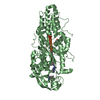
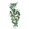
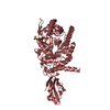
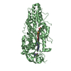
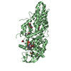
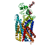
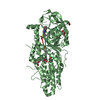
 Structure visualization
 Downloads & links
 Other downloads
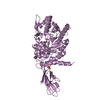


 PDBj
PDBj
 Assembly


 Type: L-peptide linking / Mass: 115.130 Da / Num. of mol.: 1 / Source method: obtained synthetically / Formula: C5H9NO2
 Mass: 24.305 Da / Num. of mol.: 1 / Source method: obtained synthetically / Formula: Mg
 Mass: 92.094 Da / Num. of mol.: 4 / Source method: obtained synthetically / Formula: C3H8O3
 Mass: 18.015 Da / Num. of mol.: 267 / Source method: isolated from a natural source / Formula: H2O
 Sample preparation
 Processing