 Movie
Movie Controller

[English] 日本語
 Yorodumi
- PDB-2x2i: Crystal structure of the Gracilariopsis lemaneiformis alpha-1,4- ... -
+ Open data

- Basic information
| Entry | Database: PDB / ID: 2x2i | |||||||||
|---|---|---|---|---|---|---|---|---|---|---|
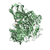
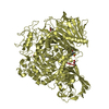
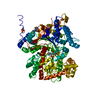
| Title | Crystal structure of the Gracilariopsis lemaneiformis alpha-1,4- glucan lyase with acarbose | |||||||||
 Components Components | ALPHA-1,4-GLUCAN LYASE ISOZYME 1 | |||||||||
 Keywords Keywords | LYASE / ANHYDROFRUCTOSE PATHWAY / GLYCOSIDE HYDROLASE FAMILY 31 / STARCH BINDING DOMAIN | |||||||||
| Function / homology |  Function and homology information Function and homology informationexo-(1->4)-alpha-D-glucan lyase / exo-(1,4)-alpha-D-glucan lyase activity / hydrolase activity, hydrolyzing O-glycosyl compounds / carbohydrate binding / carbohydrate metabolic process Similarity search - Function | |||||||||
| Biological species |  GRACILARIOPSIS LEMANEIFORMIS (eukaryote) GRACILARIOPSIS LEMANEIFORMIS (eukaryote) | |||||||||
| Method |  X-RAY DIFFRACTION / MOLECULAR REPLACEMENT / Resolution: 2.6 Å X-RAY DIFFRACTION / MOLECULAR REPLACEMENT / Resolution: 2.6 Å | |||||||||
 Authors Authors | Rozeboom, H.J. / Yu, S. / Madrid, S. / Kalk, K.H. / Dijkstra, B.W. | |||||||||
 Citation Citation | Journal: J.Biol.Chem. / Year: 2013 Title: Crystal Structure of Alpha-1,4-Glucan Lyase, a Unique Glycoside Hydrolase Family Member with a Novel Catalytic Mechanism. Authors: Rozeboom, H.J. / Yu, S. / Madrid, S. / Kalk, K.H. / Zhang, R. / Dijkstra, B.W. | |||||||||
| History |
|
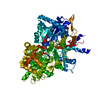
- Structure visualization
| Structure viewer | Molecule: MolmilJmol/JSmol |
|---|
- Downloads & links
-Download
| PDBx/mmCIF format | 2x2i.cif.gz | 828.9 KB | Display | PDBx/mmCIF format |
|---|---|---|---|---|
| PDB format | pdb2x2i.ent.gz | 670.7 KB | Display | PDB format |
| PDBx/mmJSON format | 2x2i.json.gz | Tree view | PDBx/mmJSON format | |
| Others |  Other downloads Other downloads |
-Validation report
| Arichive directory | https://data.pdbj.org/pub/pdb/validation_reports/x2/2x2iftp://data.pdbj.org/pub/pdb/validation_reports/x2/2x2i | HTTPS FTP |
|---|
-Related structure data
| Related structure data |  2x2hSC  2x2jC 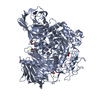 4amwC 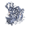 4amxC S: Starting model for refinement C: citing same article ( |
|---|---|
| Similar structure data |


-Links
 PDBj
PDBj


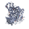
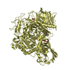
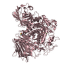
- Assembly
| Deposited unit | 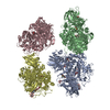
| ||||||||||||||||||||||||||||||
|---|---|---|---|---|---|---|---|---|---|---|---|---|---|---|---|---|---|---|---|---|---|---|---|---|---|---|---|---|---|---|---|
| 1 | 
| ||||||||||||||||||||||||||||||
| 2 | 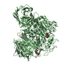
| ||||||||||||||||||||||||||||||
| 3 | 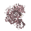
| ||||||||||||||||||||||||||||||
| 4 | 
| ||||||||||||||||||||||||||||||
| Unit cell |
| ||||||||||||||||||||||||||||||
| Noncrystallographic symmetry (NCS) | NCS domain:
NCS domain segments: Component-ID: 1 / Ens-ID: 1 / Beg auth comp-ID: THR / Beg label comp-ID: THR / End auth comp-ID: THR / End label comp-ID: THR / Refine code: 5 / Auth seq-ID: 14 - 1038 / Label seq-ID: 3 - 1027
|
-Components
| #1: Protein | Mass: 116062.156 Da / Num. of mol.: 4 / Fragment: RESIDUES 62-1088 Source method: isolated from a genetically manipulated source Source: (gene. exp.) GRACILARIOPSIS LEMANEIFORMIS (eukaryote)Description: REPLICATING VECTOR WITH HARS SEQUENCE. COLLECTED AT TAPING BAY, TSINGTAO, CHINA Production host:  PICHIA ANGUSTA (fungus) / Strain (production host): RB11 PICHIA ANGUSTA (fungus) / Strain (production host): RB11References: UniProt: Q9STC1, exo-(1->4)-alpha-D-glucan lyase #2: Polysaccharide | 4,6-dideoxy-4-{[(1S,4R,5S,6S)-4,5,6-trihydroxy-3-(hydroxymethyl)cyclohex-2-en-1-yl]amino}-alpha-D- ...4,6-dideoxy-4-{[(1S,4R,5S,6S)-4,5,6-trihydroxy-3-(hydroxymethyl)cyclohex-2-en-1-yl]amino}-alpha-D-glucopyranose-(1-4)-alpha-D-glucopyranose-(1-4)-beta-D-glucopyranose / beta-acarbose 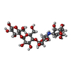  Type: oligosaccharide, Oligosaccharide / Class: Inhibitor / Mass: 645.606 Da / Num. of mol.: 8 Type: oligosaccharide, Oligosaccharide / Class: Inhibitor / Mass: 645.606 Da / Num. of mol.: 8Source method: isolated from a genetically manipulated source Details: oligosaccharide / References: beta-acarbose #3: Chemical | ChemComp-GOL /   Mass: 92.094 Da / Num. of mol.: 7 / Source method: obtained synthetically / Formula: C3H8O3 Mass: 92.094 Da / Num. of mol.: 7 / Source method: obtained synthetically / Formula: C3H8O3#4: Water | ChemComp-HOH / |  Mass: 18.015 Da / Num. of mol.: 836 / Source method: isolated from a natural source / Formula: H2O Mass: 18.015 Da / Num. of mol.: 836 / Source method: isolated from a natural source / Formula: H2OHas protein modification | Y | Sequence details | FIRST 50 AMINO ACIDS ARE A SIGNAL PEPTIDE. THE NEXT 11 AMINO ACIDS ARE NOT PRESENT IN THE ENZYME. | |
|---|
-Experimental details
-Experiment
| Experiment | Method: X-RAY DIFFRACTION / Number of used crystals: 1 |
|---|
- Sample preparation
| Crystal | Density Matthews: 2.5 Å3/Da / Density % sol: 51 % / Description: NONE |
|---|---|
| Crystal grow | Temperature: 298 K / Method: vapor diffusion, hanging drop / pH: 5 Details: 18-21% PEG 8000, 0.1M SODIUM ACETATE, PH 5.0-5.2, VAPOR DIFFUSION, HANGING DROP, TEMPERATURE 298K |
-Data collection
| Diffraction | Mean temperature: 112 K |
|---|---|
| Diffraction source | Source: ROTATING ANODE / Type: ENRAF-NONIUS FR591 / Wavelength: 1.5418 |
| Detector | Type: MACSCIENCE DIP2030H / Detector: IMAGE PLATE / Date: Oct 12, 2006 / Details: FRANKS MIRRORS |
| Radiation | Protocol: SINGLE WAVELENGTH / Monochromatic (M) / Laue (L): M / Scattering type: x-ray |
| Radiation wavelength | Wavelength: 1.5418 Å / Relative weight: 1 |
| Reflection | Resolution: 2.6→50 Å / Num. obs: 118187 / % possible obs: 82.8 % / Observed criterion σ(I): -3 / Redundancy: 2.7 % / Rmerge(I) obs: 0.23 / Net I/σ(I): 4.6 |
| Reflection shell | Resolution: 2.6→2.69 Å / Redundancy: 2.1 % / Rmerge(I) obs: 0.64 / Mean I/σ(I) obs: 1 / % possible all: 79.3 |
- Processing
| Software |
| ||||||||||||||||||||||||||||||||||||||||||||||||||||||||||||||||||||||||||||||||||||||||||||||||||||||||||||||||||||||||||||||||||||||||||||||||||||||||||||||||||||||||||||||||||||||
|---|---|---|---|---|---|---|---|---|---|---|---|---|---|---|---|---|---|---|---|---|---|---|---|---|---|---|---|---|---|---|---|---|---|---|---|---|---|---|---|---|---|---|---|---|---|---|---|---|---|---|---|---|---|---|---|---|---|---|---|---|---|---|---|---|---|---|---|---|---|---|---|---|---|---|---|---|---|---|---|---|---|---|---|---|---|---|---|---|---|---|---|---|---|---|---|---|---|---|---|---|---|---|---|---|---|---|---|---|---|---|---|---|---|---|---|---|---|---|---|---|---|---|---|---|---|---|---|---|---|---|---|---|---|---|---|---|---|---|---|---|---|---|---|---|---|---|---|---|---|---|---|---|---|---|---|---|---|---|---|---|---|---|---|---|---|---|---|---|---|---|---|---|---|---|---|---|---|---|---|---|---|---|---|
| Refinement | Method to determine structure: MOLECULAR REPLACEMENT Starting model: PDB ENTRY 2X2H Resolution: 2.6→41.85 Å / Cor.coef. Fo:Fc: 0.941 / Cor.coef. Fo:Fc free: 0.875 / SU B: 17.369 / SU ML: 0.358 / Cross valid method: THROUGHOUT / ESU R Free: 0.497 / Stereochemistry target values: MAXIMUM LIKELIHOOD
| ||||||||||||||||||||||||||||||||||||||||||||||||||||||||||||||||||||||||||||||||||||||||||||||||||||||||||||||||||||||||||||||||||||||||||||||||||||||||||||||||||||||||||||||||||||||
| Solvent computation | Ion probe radii: 0.8 Å / Shrinkage radii: 0.8 Å / VDW probe radii: 1.4 Å / Solvent model: BABINET MODEL WITH MASK | ||||||||||||||||||||||||||||||||||||||||||||||||||||||||||||||||||||||||||||||||||||||||||||||||||||||||||||||||||||||||||||||||||||||||||||||||||||||||||||||||||||||||||||||||||||||
| Displacement parameters | Biso mean: 40.005 Å2
| ||||||||||||||||||||||||||||||||||||||||||||||||||||||||||||||||||||||||||||||||||||||||||||||||||||||||||||||||||||||||||||||||||||||||||||||||||||||||||||||||||||||||||||||||||||||
| Refinement step | Cycle: LAST / Resolution: 2.6→41.85 Å
| ||||||||||||||||||||||||||||||||||||||||||||||||||||||||||||||||||||||||||||||||||||||||||||||||||||||||||||||||||||||||||||||||||||||||||||||||||||||||||||||||||||||||||||||||||||||
| Refine LS restraints |
|