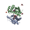
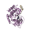
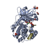
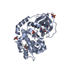
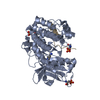
[English] 日本語
Yorodumi
- PDB-2wtg: High resolution 3D structure of C.elegans globin-like protein GLB-1 -

+
Open data


ID or keywords:

Loading...

-
Basic information

Entry
Database: PDB / ID: 2wtg
TitleHigh resolution 3D structure of C.elegans globin-like protein GLB-1
ComponentsGLOBIN-LIKE PROTEIN
KeywordsOXYGEN TRANSPORT / METAL-BINDING
Function / homology
Function and homology information


oxygen carrier activity / oxygen binding / iron ion binding / heme binding / cytoplasm
Similarity search - Function
Globin, nematode / Myoglobin-like, M family globin domain / Globin/Protoglobin / Globins / Globin domain profile. / Globin-like / Globin / Globin / Globin-like superfamily / Orthogonal Bundle / Mainly Alpha
Similarity search - Domain/homology
PROTOPORPHYRIN IX CONTAINING FE / OXYGEN MOLECULE / Globin-like protein
Similarity search - Component
Biological speciesCAENORHABDITIS ELEGANS (invertebrata)
MethodX-RAY DIFFRACTION / SYNCHROTRON / MAD / Resolution: 1.5 Å
AuthorsGeuens, E. / Hoogewijs, D. / Nardini, M. / Vinck, E. / Pesce, A. / Kiger, L. / Fago, A. / Tilleman, L. / De Henau, S. / Marden, M. ...Geuens, E. / Hoogewijs, D. / Nardini, M. / Vinck, E. / Pesce, A. / Kiger, L. / Fago, A. / Tilleman, L. / De Henau, S. / Marden, M. / Weber, R.E. / Van Doorslaer, S. / Vanfleteren, J. / Moens, L. / Bolognesi, M. / Dewilde, S.
CitationJournal: Bmc Biochem. / Year: 2010
Title: Globin-Like Proteins in Caenorhabditis Elegans: In Vivo Localization, Ligand Binding and Structural Properties.
Authors: Geuens, E. / Hoogewijs, D. / Nardini, M. / Vinck, E. / Pesce, A. / Kiger, L. / Fago, A. / Tilleman, L. / De Henau, S. / Marden, M. / Weber, R.E. / Van Doorslaer, S. / Vanfleteren, J. / ...Authors: Geuens, E. / Hoogewijs, D. / Nardini, M. / Vinck, E. / Pesce, A. / Kiger, L. / Fago, A. / Tilleman, L. / De Henau, S. / Marden, M. / Weber, R.E. / Van Doorslaer, S. / Vanfleteren, J. / Moens, L. / Bolognesi, M. / De Wilde, S.
History
DepositionSep 16, 2009Deposition site: PDBE / Processing site: PDBE
Revision 1.0Apr 21, 2010Provider: repository / Type: Initial release
Revision 1.1May 8, 2011Group: Version format compliance
Revision 1.2Jul 13, 2011Group: Version format compliance
Revision 1.3May 8, 2024Group: Data collection / Database references ...Data collection / Database references / Derived calculations / Other
Category: chem_comp_atom / chem_comp_bond ...chem_comp_atom / chem_comp_bond / database_2 / pdbx_database_status / pdbx_struct_conn_angle / struct_conn / struct_site
Item: _database_2.pdbx_DOI / _database_2.pdbx_database_accession ..._database_2.pdbx_DOI / _database_2.pdbx_database_accession / _pdbx_database_status.status_code_sf / _pdbx_struct_conn_angle.ptnr1_auth_comp_id / _pdbx_struct_conn_angle.ptnr1_auth_seq_id / _pdbx_struct_conn_angle.ptnr1_label_asym_id / _pdbx_struct_conn_angle.ptnr1_label_atom_id / _pdbx_struct_conn_angle.ptnr1_label_comp_id / _pdbx_struct_conn_angle.ptnr1_label_seq_id / _pdbx_struct_conn_angle.ptnr3_auth_comp_id / _pdbx_struct_conn_angle.ptnr3_auth_seq_id / _pdbx_struct_conn_angle.ptnr3_label_asym_id / _pdbx_struct_conn_angle.ptnr3_label_atom_id / _pdbx_struct_conn_angle.ptnr3_label_comp_id / _pdbx_struct_conn_angle.ptnr3_label_seq_id / _pdbx_struct_conn_angle.value / _struct_conn.pdbx_dist_value / _struct_conn.ptnr1_auth_comp_id / _struct_conn.ptnr1_auth_seq_id / _struct_conn.ptnr1_label_asym_id / _struct_conn.ptnr1_label_atom_id / _struct_conn.ptnr1_label_comp_id / _struct_conn.ptnr1_label_seq_id / _struct_conn.ptnr2_auth_comp_id / _struct_conn.ptnr2_auth_seq_id / _struct_conn.ptnr2_label_asym_id / _struct_conn.ptnr2_label_atom_id / _struct_conn.ptnr2_label_comp_id / _struct_conn.ptnr2_label_seq_id / _struct_site.pdbx_auth_asym_id / _struct_site.pdbx_auth_comp_id / _struct_site.pdbx_auth_seq_id

-
Structure visualization

Structure viewerMolecule:
MolmilJmol/JSmol

Downloads & links

-
Assembly

Deposited unit
A: GLOBIN-LIKE PROTEIN
hetero molecules


Theoretical massNumber of molelcules
Total (without water)19,1573
Polymers18,5091
Non-polymers6482
Water3,675204
1
A: GLOBIN-LIKE PROTEIN
hetero molecules

A: GLOBIN-LIKE PROTEIN
hetero molecules


Theoretical massNumber of molelcules
Total (without water)38,3156
Polymers37,0182
Non-polymers1,2974
Water362
TypeNameSymmetry operationNumber
identity operation1_555x,y,z1
crystal symmetry operation7_555y,x,-z1
Buried area4620 Å2
ΔGint-55.6 kcal/mol
Surface area14660 Å2
MethodPISA
Unit cell
Length a, b, c (Å)81.947, 81.947, 47.042
Angle α, β, γ (deg.)90.00, 90.00, 90.00
Int Tables number96
Space group name H-MP43212
Components on special symmetry positions
IDModelComponents
11A-2028-

HOH

21A-2150-

HOH

31A-2151-

HOH

-
Components

#1: Protein GLOBIN-LIKE PROTEIN


Mass: 18508.979 Da / Num. of mol.: 1 / Mutation: YES
Source method: isolated from a genetically manipulated source
Source: (gene. exp.) CAENORHABDITIS ELEGANS (invertebrata) / Production host: ESCHERICHIA COLI (E. coli) / References: UniProt: P30627
#2: Chemical ChemComp-HEM / PROTOPORPHYRIN IX CONTAINING FE / HEME


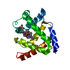
Mass: 616.487 Da / Num. of mol.: 1 / Source method: obtained synthetically / Formula: C34H32FeN4O4
#3: Chemical ChemComp-OXY / OXYGEN MOLECULE


Mass: 31.999 Da / Num. of mol.: 1 / Source method: obtained synthetically / Formula: O2
#4: Water ChemComp-HOH / water


Mass: 18.015 Da / Num. of mol.: 204 / Source method: isolated from a natural source / Formula: H2O
Compound detailsENGINEERED RESIDUE IN CHAIN A, CYS 127 TO SER
Sequence detailsWITH RESPECT TO SEQUENCE PRESENT IN THE UNIPROT DATABASE, CYS127 WAS MUTATED INTO SER RESIDUE FOR ...WITH RESPECT TO SEQUENCE PRESENT IN THE UNIPROT DATABASE, CYS127 WAS MUTATED INTO SER RESIDUE FOR CRYSTALLIZATION PURPOSE

-
Experimental details

-
Experiment

ExperimentMethod: X-RAY DIFFRACTION / Number of used crystals: 1

-
Sample preparation

CrystalDensity Matthews: 2.2 Å3/Da / Density % sol: 44.3 % / Description: NONE
Crystal growpH: 5.5 / Details: PEG 4K 10%, 0.1 M SODIUM ACETATE PH 5.5

-
Data collection

DiffractionMean temperature: 100 K
Diffraction sourceSource: SYNCHROTRON / Site: ESRF / Beamline: ID29 / Wavelength: 0.976
DetectorType: ADSC CCD / Detector: CCD / Date: Jan 22, 2007
RadiationProtocol: MAD / Monochromatic (M) / Laue (L): M / Scattering type: x-ray
Radiation wavelengthWavelength: 0.976 Å / Relative weight: 1
ReflectionResolution: 1.5→36.6 Å / Num. obs: 25735 / % possible obs: 98.1 % / Observed criterion σ(I): 2 / Redundancy: 28.1 % / Rmerge(I) obs: 0.1 / Net I/σ(I): 30.7

-
Processing

Software
NameVersionClassification
REFMAC5.2.0019refinement
MOSFLMdata reduction
SCALAdata scaling
SOLVEphasing
RefinementMethod to determine structure: MAD
Starting model: NONE

Resolution: 1.5→36.64 Å / Cor.coef. Fo:Fc: 0.959 / Cor.coef. Fo:Fc free: 0.948 / SU B: 2.257 / SU ML: 0.04 / Cross valid method: THROUGHOUT / ESU R: 0.106 / ESU R Free: 0.082 / Stereochemistry target values: MAXIMUM LIKELIHOOD / Details: HYDROGENS HAVE BEEN ADDED IN THE RIDING POSITIONS.
RfactorNum. reflection% reflectionSelection details
Rfree0.20118 2584 10.1 %RANDOM
Rwork0.16318 ---
obs0.16699 23035 97.71 %-
Solvent computationIon probe radii: 0.8 Å / Shrinkage radii: 0.8 Å / VDW probe radii: 1.2 Å / Solvent model: MASK
Displacement parametersBiso mean: 14.625 Å2
Baniso -1Baniso -2Baniso -3
1-0 Å20 Å20 Å2
2--0 Å20 Å2
3---0 Å2
Refinement stepCycle: LAST / Resolution: 1.5→36.64 Å
ProteinNucleic acidLigandSolventTotal
Num. atoms1294 0 45 204 1543
Refine LS restraints
Refine-IDTypeDev idealDev ideal targetNumber
X-RAY DIFFRACTIONr_bond_refined_d0.0120.0221387
X-RAY DIFFRACTIONr_bond_other_d
X-RAY DIFFRACTIONr_angle_refined_deg1.4312.031884
X-RAY DIFFRACTIONr_angle_other_deg
X-RAY DIFFRACTIONr_dihedral_angle_1_deg6.455161
X-RAY DIFFRACTIONr_dihedral_angle_2_deg34.59723.83673
X-RAY DIFFRACTIONr_dihedral_angle_3_deg12.3315236
X-RAY DIFFRACTIONr_dihedral_angle_4_deg8.8491510
X-RAY DIFFRACTIONr_chiral_restr0.1010.2187
X-RAY DIFFRACTIONr_gen_planes_refined0.0080.021083
X-RAY DIFFRACTIONr_gen_planes_other
X-RAY DIFFRACTIONr_nbd_refined0.2740.2735
X-RAY DIFFRACTIONr_nbd_other
X-RAY DIFFRACTIONr_nbtor_refined0.3130.2976
X-RAY DIFFRACTIONr_nbtor_other
X-RAY DIFFRACTIONr_xyhbond_nbd_refined0.1520.2160
X-RAY DIFFRACTIONr_xyhbond_nbd_other
X-RAY DIFFRACTIONr_metal_ion_refined
X-RAY DIFFRACTIONr_metal_ion_other
X-RAY DIFFRACTIONr_symmetry_vdw_refined0.1860.232
X-RAY DIFFRACTIONr_symmetry_vdw_other
X-RAY DIFFRACTIONr_symmetry_hbond_refined0.1220.223
X-RAY DIFFRACTIONr_symmetry_hbond_other
X-RAY DIFFRACTIONr_symmetry_metal_ion_refined
X-RAY DIFFRACTIONr_symmetry_metal_ion_other
X-RAY DIFFRACTIONr_mcbond_it1.0681.5802
X-RAY DIFFRACTIONr_mcbond_other
X-RAY DIFFRACTIONr_mcangle_it1.60421268
X-RAY DIFFRACTIONr_mcangle_other
X-RAY DIFFRACTIONr_scbond_it2.4473662
X-RAY DIFFRACTIONr_scbond_other
X-RAY DIFFRACTIONr_scangle_it3.4384.5612
X-RAY DIFFRACTIONr_scangle_other
X-RAY DIFFRACTIONr_long_range_B_refined
X-RAY DIFFRACTIONr_long_range_B_other
X-RAY DIFFRACTIONr_rigid_bond_restr1.41731464
X-RAY DIFFRACTIONr_sphericity_free3.823206
X-RAY DIFFRACTIONr_sphericity_bonded2.57231348
LS refinement shellResolution: 1.5→1.539 Å / Total num. of bins used: 20
RfactorNum. reflection% reflection
Rfree0.216 188 -
Rwork0.136 1652 -
obs--96.34 %

+
About Yorodumi

-
News

-
Feb 9, 2022. New format data for meta-information of EMDB entries

New format data for meta-information of EMDB entries

  • Version 3 of the EMDB header file is now the official format.
  • The previous official version 1.9 will be removed from the archive.

Related info.:EMDB header

External links:wwPDB to switch to version 3 of the EMDB data model

-
Aug 12, 2020. Covid-19 info

Covid-19 info

URL: https://pdbjlvh1.pdbj.org/emnavi/covid19.php

New page: Covid-19 featured information page in EM Navigator.

Related info.:Covid-19 info / Mar 5, 2020. Novel coronavirus structure data

+
Mar 5, 2020. Novel coronavirus structure data

Novel coronavirus structure data

Related info.:Yorodumi Speices / Aug 12, 2020. Covid-19 info

External links:COVID-19 featured content - PDBj / Molecule of the Month (242):Coronavirus Proteases

+
Jan 31, 2019. EMDB accession codes are about to change! (news from PDBe EMDB page)

EMDB accession codes are about to change! (news from PDBe EMDB page)

  • The allocation of 4 digits for EMDB accession codes will soon come to an end. Whilst these codes will remain in use, new EMDB accession codes will include an additional digit and will expand incrementally as the available range of codes is exhausted. The current 4-digit format prefixed with “EMD-” (i.e. EMD-XXXX) will advance to a 5-digit format (i.e. EMD-XXXXX), and so on. It is currently estimated that the 4-digit codes will be depleted around Spring 2019, at which point the 5-digit format will come into force.
  • The EM Navigator/Yorodumi systems omit the EMD- prefix.

Related info.:Q: What is EMD? / ID/Accession-code notation in Yorodumi/EM Navigator

External links:EMDB Accession Codes are Changing Soon! / Contact to PDBj

+
Jul 12, 2017. Major update of PDB

Major update of PDB

  • wwPDB released updated PDB data conforming to the new PDBx/mmCIF dictionary.
  • This is a major update changing the version number from 4 to 5, and with Remediation, in which all the entries are updated.
  • In this update, many items about electron microscopy experimental information are reorganized (e.g. em_software).
  • Now, EM Navigator and Yorodumi are based on the updated data.

External links:wwPDB Remediation / Enriched Model Files Conforming to OneDep Data Standards Now Available in the PDB FTP Archive

-
Yorodumi

Thousand views of thousand structures

  • Yorodumi is a browser for structure data from EMDB, PDB, SASBDB, etc.
  • This page is also the successor to EM Navigator detail page, and also detail information page/front-end page for Omokage search.
  • The word "yorodu" (or yorozu) is an old Japanese word meaning "ten thousand". "mi" (miru) is to see.

Related info.:EMDB / PDB / SASBDB / Comparison of 3 databanks / Yorodumi Search / Aug 31, 2016. New EM Navigator & Yorodumi / Yorodumi Papers / Jmol/JSmol / Function and homology information / Changes in new EM Navigator and Yorodumi

Read more