[English] 日本語
Yorodumi
- PDB-2wjh: Structure and function of the FeoB G-domain from Methanococcus ja... -

+
Open data


ID or keywords:

Loading...

-
Basic information

Entry
Database: PDB / ID: 2wjh
Title
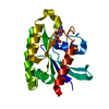
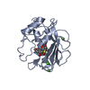
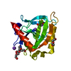
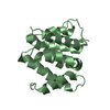
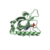
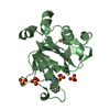
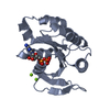
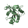
Structure and function of the FeoB G-domain from Methanococcus jannaschii
ComponentsFERROUS IRON TRANSPORT PROTEIN B HOMOLOG
KeywordsMETAL TRANSPORT / MEMBRANE G-PROTEINS / FERROUS IRON TRANSPORT / CELL MEMBRANE / ION TRANSPORT / TRANSMEMBRANE / NUCLEOTIDE BINDING MOTIFS / IRON / GNBPS / MEMBRANE / TRANSPORT / GTP-BINDING / IRON TRANSPORT / NUCLEOTIDE-BINDING
Function / homology
Function and homology information


ferrous iron transmembrane transporter activity / GTP binding / metal ion binding / plasma membrane
Similarity search - Function
FeoB, cytosolic helical domain / FeoB cytosolic helical domain / Ferrous iron transport protein B, C-terminal / Ferrous iron transport protein B C terminus / Ferrous iron transport protein B / FeoB-type guanine nucleotide-binding (G) domain / Ferrous iron transport protein B / FeoB-type guanine nucleotide-binding (G) domain profile. / Nucleoside transporter/FeoB GTPase, Gate domain / Nucleoside recognition ...FeoB, cytosolic helical domain / FeoB cytosolic helical domain / Ferrous iron transport protein B, C-terminal / Ferrous iron transport protein B C terminus / Ferrous iron transport protein B / FeoB-type guanine nucleotide-binding (G) domain / Ferrous iron transport protein B / FeoB-type guanine nucleotide-binding (G) domain profile. / Nucleoside transporter/FeoB GTPase, Gate domain / Nucleoside recognition / GTP binding domain / Small GTP-binding protein domain / P-loop containing nucleotide triphosphate hydrolases / P-loop containing nucleoside triphosphate hydrolase / Rossmann fold / 3-Layer(aba) Sandwich / Alpha Beta
Similarity search - Domain/homology
CITRATE ANION / GUANOSINE-5'-DIPHOSPHATE / Fe(2+) transporter FeoB
Similarity search - Component
Biological speciesMETHANOCALDOCOCCUS JANNASCHII (archaea)
MethodX-RAY DIFFRACTION / SYNCHROTRON / MOLECULAR REPLACEMENT / Resolution: 2.1 Å
AuthorsKoester, S. / Wehner, M. / Herrmann, C. / Kuehlbrandt, W. / Yildiz, O.
CitationJournal: J.Mol.Biol. / Year: 2009
Title: Structure and Function of the Feob G-Domain from Methanococcus Jannaschii
Authors: Koester, S. / Wehner, M. / Herrmann, C. / Kuehlbrandt, W. / Yildiz, O.
History
DepositionMay 26, 2009Deposition site: PDBE / Processing site: PDBE
Revision 1.0Jul 28, 2009Provider: repository / Type: Initial release
Revision 1.1May 19, 2011Group: Version format compliance
Revision 1.2Jul 13, 2011Group: Version format compliance
Revision 1.3Dec 13, 2023Group: Data collection / Database references ...Data collection / Database references / Derived calculations / Other / Refinement description
Category: chem_comp_atom / chem_comp_bond ...chem_comp_atom / chem_comp_bond / database_2 / pdbx_database_status / pdbx_initial_refinement_model / pdbx_struct_conn_angle / struct_conn / struct_site
Item: _database_2.pdbx_DOI / _database_2.pdbx_database_accession ..._database_2.pdbx_DOI / _database_2.pdbx_database_accession / _pdbx_database_status.status_code_sf / _pdbx_struct_conn_angle.ptnr1_auth_comp_id / _pdbx_struct_conn_angle.ptnr1_auth_seq_id / _pdbx_struct_conn_angle.ptnr1_label_asym_id / _pdbx_struct_conn_angle.ptnr1_label_atom_id / _pdbx_struct_conn_angle.ptnr1_label_comp_id / _pdbx_struct_conn_angle.ptnr1_label_seq_id / _pdbx_struct_conn_angle.ptnr3_auth_comp_id / _pdbx_struct_conn_angle.ptnr3_auth_seq_id / _pdbx_struct_conn_angle.ptnr3_label_asym_id / _pdbx_struct_conn_angle.ptnr3_label_atom_id / _pdbx_struct_conn_angle.ptnr3_label_comp_id / _pdbx_struct_conn_angle.ptnr3_label_seq_id / _pdbx_struct_conn_angle.value / _struct_conn.pdbx_dist_value / _struct_conn.ptnr1_auth_comp_id / _struct_conn.ptnr1_auth_seq_id / _struct_conn.ptnr1_label_asym_id / _struct_conn.ptnr1_label_atom_id / _struct_conn.ptnr1_label_comp_id / _struct_conn.ptnr1_label_seq_id / _struct_conn.ptnr2_auth_comp_id / _struct_conn.ptnr2_auth_seq_id / _struct_conn.ptnr2_label_asym_id / _struct_conn.ptnr2_label_atom_id / _struct_conn.ptnr2_label_comp_id / _struct_conn.ptnr2_label_seq_id / _struct_site.pdbx_auth_asym_id / _struct_site.pdbx_auth_comp_id / _struct_site.pdbx_auth_seq_id

-
Structure visualization

Structure viewerMolecule:
MolmilJmol/JSmol

Downloads & links

-
Assembly

Deposited unit
A: FERROUS IRON TRANSPORT PROTEIN B HOMOLOG
B: FERROUS IRON TRANSPORT PROTEIN B HOMOLOG
hetero molecules


Theoretical massNumber of molelcules
Total (without water)37,1967
Polymers36,4912
Non-polymers7055
Water3,117173
1
A: FERROUS IRON TRANSPORT PROTEIN B HOMOLOG
hetero molecules


Theoretical massNumber of molelcules
Total (without water)18,7374
Polymers18,2451
Non-polymers4923
Water181
TypeNameSymmetry operationNumber
identity operation1_555x,y,z1
MethodPISA
2
B: FERROUS IRON TRANSPORT PROTEIN B HOMOLOG
hetero molecules


Theoretical massNumber of molelcules
Total (without water)18,4593
Polymers18,2451
Non-polymers2132
Water181
TypeNameSymmetry operationNumber
identity operation1_555x,y,z1
MethodPISA
Unit cell
Length a, b, c (Å)104.200, 50.400, 94.700
Angle α, β, γ (deg.)90.00, 120.60, 90.00
Int Tables number5
Space group name H-MC121

-
Components

#1: Protein FERROUS IRON TRANSPORT PROTEIN B HOMOLOG / FEOB


Mass: 18245.260 Da / Num. of mol.: 2 / Fragment: FEOB G-DOMAIN, RESIDUES 1-165
Source method: isolated from a genetically manipulated source
Source: (gene. exp.) METHANOCALDOCOCCUS JANNASCHII (archaea)
Plasmid: PET / Production host: ESCHERICHIA COLI (E. coli) / References: UniProt: Q57986
#2: Chemical ChemComp-GDP / GUANOSINE-5'-DIPHOSPHATE


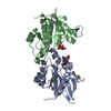
Type: RNA linking / Mass: 443.201 Da / Num. of mol.: 1 / Source method: obtained synthetically / Formula: C10H15N5O11P2 / Comment: GDP, energy-carrying molecule*YM
#3: Chemical ChemComp-MG / MAGNESIUM ION


Mass: 24.305 Da / Num. of mol.: 3 / Source method: obtained synthetically / Formula: Mg
#4: Chemical ChemComp-FLC / CITRATE ANION


Mass: 189.100 Da / Num. of mol.: 1 / Source method: obtained synthetically / Formula: C6H5O7
#5: Water ChemComp-HOH / water


Mass: 18.015 Da / Num. of mol.: 173 / Source method: isolated from a natural source / Formula: H2O

-
Experimental details

-
Experiment

ExperimentMethod: X-RAY DIFFRACTION / Number of used crystals: 1

-
Sample preparation

CrystalDensity Matthews: 2.75 Å3/Da / Density % sol: 54.9 % / Description: NONE

-
Data collection

DiffractionMean temperature: 100 K
Diffraction sourceSource: SYNCHROTRON / Site: ESRF / Beamline: ID14-4 / Wavelength: 0.9395
DetectorType: ADSC CCD / Detector: CCD / Date: Feb 8, 2008
RadiationProtocol: SINGLE WAVELENGTH / Monochromatic (M) / Laue (L): M / Scattering type: x-ray
Radiation wavelengthWavelength: 0.9395 Å / Relative weight: 1
ReflectionResolution: 2.1→20 Å / Num. obs: 24931 / % possible obs: 99.6 % / Observed criterion σ(I): 2.9 / Redundancy: 10.2 % / Biso Wilson estimate: 31.64 Å2 / Rmerge(I) obs: 0.08 / Net I/σ(I): 14.5
Reflection shellResolution: 2.1→2.2 Å / Redundancy: 5.5 % / Rmerge(I) obs: 0.55 / Mean I/σ(I) obs: 2.9 / % possible all: 99.7

-
Processing

Software
NameVersionClassification
PHENIX(PHENIX.REFINE)refinement
XDSdata reduction
XSCALEdata scaling
PHASERphasing
RefinementMethod to determine structure: MOLECULAR REPLACEMENT
Starting model: PDB ENTRY 2WJG
Resolution: 2.1→42.944 Å / SU ML: 0.37 / σ(F): 2.02 / Phase error: 24.35 / Stereochemistry target values: ML
RfactorNum. reflection% reflection
Rfree0.2461 1247 5 %
Rwork0.2104 --
obs0.2122 24927 99.9 %
Solvent computationShrinkage radii: 0.9 Å / VDW probe radii: 1.11 Å / Solvent model: FLAT BULK SOLVENT MODEL / Bsol: 60.836 Å2 / ksol: 0.347 e/Å3
Displacement parameters
Baniso -1Baniso -2Baniso -3
1--5.5582 Å20 Å2-7.2549 Å2
2---0.2243 Å2-0 Å2
3---5.7824 Å2
Refinement stepCycle: LAST / Resolution: 2.1→42.944 Å
ProteinNucleic acidLigandSolventTotal
Num. atoms2558 0 44 173 2775
Refine LS restraints
Refine-IDTypeDev idealNumber
X-RAY DIFFRACTIONf_bond_d0.0082637
X-RAY DIFFRACTIONf_angle_d1.1153570
X-RAY DIFFRACTIONf_dihedral_angle_d18.788992
X-RAY DIFFRACTIONf_chiral_restr0.065430
X-RAY DIFFRACTIONf_plane_restr0.004443
LS refinement shell
Resolution (Å)Rfactor RfreeNum. reflection RfreeRfactor RworkNum. reflection RworkRefine-ID% reflection obs (%)
2.1-2.18410.28061370.24572601X-RAY DIFFRACTION100
2.1841-2.28350.28241370.23072614X-RAY DIFFRACTION100
2.2835-2.40390.28771380.22582618X-RAY DIFFRACTION100
2.4039-2.55440.2891370.22022601X-RAY DIFFRACTION100
2.5544-2.75160.25681390.22082630X-RAY DIFFRACTION100
2.7516-3.02850.25571370.22472612X-RAY DIFFRACTION100
3.0285-3.46650.26571390.21162644X-RAY DIFFRACTION100
3.4665-4.36680.21061400.18322647X-RAY DIFFRACTION100
4.3668-42.95280.20791430.17942713X-RAY DIFFRACTION99
Refinement TLS params.Method: refined / Origin x: -23.256 Å / Origin y: 9.5226 Å / Origin z: -18.5593 Å
111213212223313233
T0.1742 Å2-0.0189 Å2-0.0091 Å2-0.1623 Å20.0009 Å2--0.1416 Å2
L1.0104 °20.2291 °20.0007 °2-0.3173 °20.1016 °2--0.5545 °2
S-0.0699 Å °0.1925 Å °-0.0403 Å °-0.0717 Å °0.0731 Å °0.0328 Å °-0.024 Å °0.0271 Å °-0.0048 Å °
Refinement TLS groupSelection details: ALL

+
About Yorodumi

-
News

-
Feb 9, 2022. New format data for meta-information of EMDB entries

New format data for meta-information of EMDB entries

  • Version 3 of the EMDB header file is now the official format.
  • The previous official version 1.9 will be removed from the archive.

Related info.:EMDB header

External links:wwPDB to switch to version 3 of the EMDB data model

-
Aug 12, 2020. Covid-19 info

Covid-19 info

URL: https://pdbjlvh1.pdbj.org/emnavi/covid19.php

New page: Covid-19 featured information page in EM Navigator.

Related info.:Covid-19 info / Mar 5, 2020. Novel coronavirus structure data

+
Mar 5, 2020. Novel coronavirus structure data

Novel coronavirus structure data

Related info.:Yorodumi Speices / Aug 12, 2020. Covid-19 info

External links:COVID-19 featured content - PDBj / Molecule of the Month (242):Coronavirus Proteases

+
Jan 31, 2019. EMDB accession codes are about to change! (news from PDBe EMDB page)

EMDB accession codes are about to change! (news from PDBe EMDB page)

  • The allocation of 4 digits for EMDB accession codes will soon come to an end. Whilst these codes will remain in use, new EMDB accession codes will include an additional digit and will expand incrementally as the available range of codes is exhausted. The current 4-digit format prefixed with “EMD-” (i.e. EMD-XXXX) will advance to a 5-digit format (i.e. EMD-XXXXX), and so on. It is currently estimated that the 4-digit codes will be depleted around Spring 2019, at which point the 5-digit format will come into force.
  • The EM Navigator/Yorodumi systems omit the EMD- prefix.

Related info.:Q: What is EMD? / ID/Accession-code notation in Yorodumi/EM Navigator

External links:EMDB Accession Codes are Changing Soon! / Contact to PDBj

+
Jul 12, 2017. Major update of PDB

Major update of PDB

  • wwPDB released updated PDB data conforming to the new PDBx/mmCIF dictionary.
  • This is a major update changing the version number from 4 to 5, and with Remediation, in which all the entries are updated.
  • In this update, many items about electron microscopy experimental information are reorganized (e.g. em_software).
  • Now, EM Navigator and Yorodumi are based on the updated data.

External links:wwPDB Remediation / Enriched Model Files Conforming to OneDep Data Standards Now Available in the PDB FTP Archive

-
Yorodumi

Thousand views of thousand structures

  • Yorodumi is a browser for structure data from EMDB, PDB, SASBDB, etc.
  • This page is also the successor to EM Navigator detail page, and also detail information page/front-end page for Omokage search.
  • The word "yorodu" (or yorozu) is an old Japanese word meaning "ten thousand". "mi" (miru) is to see.

Related info.:EMDB / PDB / SASBDB / Comparison of 3 databanks / Yorodumi Search / Aug 31, 2016. New EM Navigator & Yorodumi / Yorodumi Papers / Jmol/JSmol / Function and homology information / Changes in new EM Navigator and Yorodumi

Read more