 Movie
Movie Controller

[English] 日本語
 Yorodumi
- PDB-2wh7: The partial structure of a group A streptpcoccal phage-encoded ta... -
+ Open data

- Basic information
| Entry | Database: PDB / ID: 2wh7 | ||||||
|---|---|---|---|---|---|---|---|
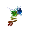
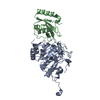
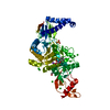
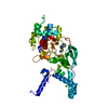
| Title | The partial structure of a group A streptpcoccal phage-encoded tail fibre hyaluronate lyase Hylp2 | ||||||
 Components Components | HYALURONIDASE-PHAGE ASSOCIATED | ||||||
 Keywords Keywords | HYDROLASE / TRIPLE-STRANDED BETA-HELIX / HYALURONAN LYASE / PHAGE TAIL FIBRE / GLYCOSIDASE / HYALURONIDASE / SCARLET FEVER | ||||||
| Function / homology | Hyaluronidase, bacterial / Hyaluronidase protein (HylP) / Major tropism determinant, N-terminal domain / Major tropism determinant N-terminal domain / hyaluronoglucosaminidase / hyalurononglucosaminidase activity / capsule polysaccharide biosynthetic process / Hyaluronidase-phage associated Function and homology information Function and homology information | ||||||
| Biological species |  STREPTOCOCCUS PYOGENES (bacteria) STREPTOCOCCUS PYOGENES (bacteria) | ||||||
| Method |  X-RAY DIFFRACTION / SYNCHROTRON / MOLECULAR REPLACEMENT / Resolution: 1.6 Å X-RAY DIFFRACTION / SYNCHROTRON / MOLECULAR REPLACEMENT / Resolution: 1.6 Å | ||||||
 Authors Authors | Martinez-Fleites, C. / Black, G.W. / Turkenburg, J.P. / Smith, N.L. / Taylor, E.J. | ||||||
 Citation Citation | Journal: Acta Crystallogr.,Sect.F / Year: 2009 Title: Structures of Two Truncated Phage-Tail Hyaluronate Lyases from Streptococcus Pyogenes Serotype M1. Authors: Martinez-Fleites, C. / Smith, N.L. / Turkenburg, J.P. / Black, G.W. / Taylor, E.J. | ||||||
| History |
|
- Structure visualization
| Structure viewer | Molecule: MolmilJmol/JSmol |
|---|
- Downloads & links
-Download
| PDBx/mmCIF format | 2wh7.cif.gz | 47.3 KB | Display | PDBx/mmCIF format |
|---|---|---|---|---|
| PDB format | pdb2wh7.ent.gz | 32.7 KB | Display | PDB format |
| PDBx/mmJSON format | 2wh7.json.gz | Tree view | PDBx/mmJSON format | |
| Others |  Other downloads Other downloads |
-Validation report
| Summary document | 2wh7_validation.pdf.gz | 420.1 KB | Display | wwPDB validaton report |
|---|---|---|---|---|
| Full document | 2wh7_full_validation.pdf.gz | 420.4 KB | Display | |
| Data in XML | 2wh7_validation.xml.gz | 10 KB | Display | |
| Data in CIF | 2wh7_validation.cif.gz | 14.5 KB | Display | |
| Arichive directory | https://data.pdbj.org/pub/pdb/validation_reports/wh/2wh7ftp://data.pdbj.org/pub/pdb/validation_reports/wh/2wh7 | HTTPS FTP |
-Related structure data
| Related structure data |  2wb3C  2c3fS S: Starting model for refinement C: citing same article ( |
|---|---|
| Similar structure data |


-Links
 PDBj
PDBj- Assembly
| Deposited unit | 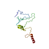
| ||||||||||||
|---|---|---|---|---|---|---|---|---|---|---|---|---|---|
| 1 | 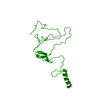
| ||||||||||||
| Unit cell |
| ||||||||||||
| Components on special symmetry positions |
|
-Components
| #1: Protein | Mass: 19118.580 Da / Num. of mol.: 1 / Fragment: HYALURONATE LYASES FRAGMENT, RESIDUES 215-372 Source method: isolated from a genetically manipulated source Source: (gene. exp.) STREPTOCOCCUS PYOGENES (bacteria) / Strain: M1 GAS SF370 / Plasmid: PET28A (NOVAGEN) / Production host: |
|---|---|
| #2: Water | ChemComp-HOH /  Mass: 18.015 Da / Num. of mol.: 200 / Source method: isolated from a natural source / Formula: H2O Mass: 18.015 Da / Num. of mol.: 200 / Source method: isolated from a natural source / Formula: H2O |
| Sequence details | THIS STRUCTURE IS A FRAGMENT, THE N-TERMINAL PORTION IS MISSING, THE STRUCTURE BEGINS AS RESIDUE 235 |
-Experimental details
-Experiment
| Experiment | Method: X-RAY DIFFRACTION / Number of used crystals: 1 |
|---|
- Sample preparation
| Crystal | Density Matthews: 2.4 Å3/Da / Density % sol: 49 % / Description: NONE |
|---|---|
| Crystal grow | Details: 2 M NACL, 10 % (W/V) PEG 6000; |
-Data collection
| Diffraction | Mean temperature: 100 K |
|---|---|
| Diffraction source | Source: SYNCHROTRON / Site: ESRF  / Beamline: ID14-1 / Wavelength: 0.933 / Beamline: ID14-1 / Wavelength: 0.933 |
| Detector | Type: ADSC CCD / Detector: CCD |
| Radiation | Protocol: SINGLE WAVELENGTH / Monochromatic (M) / Laue (L): M / Scattering type: x-ray |
| Radiation wavelength | Wavelength: 0.933 Å / Relative weight: 1 |
| Reflection | Resolution: 1.6→30 Å / Num. obs: 23240 / % possible obs: 91 % / Observed criterion σ(I): 2 / Redundancy: 4.4 % / Rmerge(I) obs: 0.07 / Net I/σ(I): 41.9 |
| Reflection shell | Resolution: 1.6→1.66 Å / Redundancy: 4.9 % / Rmerge(I) obs: 0.17 / Mean I/σ(I) obs: 13 / % possible all: 93 |
- Processing
| Software |
| ||||||||||||||||||||||||||||||||||||||||||||||||||||||||||||||||||||||||||||||||||||||||||||||||||||||||||||||||||||||||||||||||||||||||||||||||||||||||||||||||||||||||||||||||||||||
|---|---|---|---|---|---|---|---|---|---|---|---|---|---|---|---|---|---|---|---|---|---|---|---|---|---|---|---|---|---|---|---|---|---|---|---|---|---|---|---|---|---|---|---|---|---|---|---|---|---|---|---|---|---|---|---|---|---|---|---|---|---|---|---|---|---|---|---|---|---|---|---|---|---|---|---|---|---|---|---|---|---|---|---|---|---|---|---|---|---|---|---|---|---|---|---|---|---|---|---|---|---|---|---|---|---|---|---|---|---|---|---|---|---|---|---|---|---|---|---|---|---|---|---|---|---|---|---|---|---|---|---|---|---|---|---|---|---|---|---|---|---|---|---|---|---|---|---|---|---|---|---|---|---|---|---|---|---|---|---|---|---|---|---|---|---|---|---|---|---|---|---|---|---|---|---|---|---|---|---|---|---|---|---|
| Refinement | Method to determine structure: MOLECULAR REPLACEMENT Starting model: PDB ENTRY 2C3F Resolution: 1.6→109.76 Å / Cor.coef. Fo:Fc: 0.946 / Cor.coef. Fo:Fc free: 0.923 / SU B: 1.945 / SU ML: 0.07 / Cross valid method: THROUGHOUT / ESU R: 0.107 / ESU R Free: 0.111 / Stereochemistry target values: MAXIMUM LIKELIHOOD Details: HYDROGENS HAVE BEEN ADDED IN THE RIDING POSITIONS. U VALUES REFINED INDIVIDUALLY
| ||||||||||||||||||||||||||||||||||||||||||||||||||||||||||||||||||||||||||||||||||||||||||||||||||||||||||||||||||||||||||||||||||||||||||||||||||||||||||||||||||||||||||||||||||||||
| Solvent computation | Ion probe radii: 0.8 Å / Shrinkage radii: 0.8 Å / VDW probe radii: 1.4 Å / Solvent model: MASK | ||||||||||||||||||||||||||||||||||||||||||||||||||||||||||||||||||||||||||||||||||||||||||||||||||||||||||||||||||||||||||||||||||||||||||||||||||||||||||||||||||||||||||||||||||||||
| Displacement parameters | Biso mean: 18.92 Å2
| ||||||||||||||||||||||||||||||||||||||||||||||||||||||||||||||||||||||||||||||||||||||||||||||||||||||||||||||||||||||||||||||||||||||||||||||||||||||||||||||||||||||||||||||||||||||
| Refinement step | Cycle: LAST / Resolution: 1.6→109.76 Å
| ||||||||||||||||||||||||||||||||||||||||||||||||||||||||||||||||||||||||||||||||||||||||||||||||||||||||||||||||||||||||||||||||||||||||||||||||||||||||||||||||||||||||||||||||||||||
| Refine LS restraints |
|