[English] 日本語
Yorodumi
- PDB-2w2b: Crystal Structure of single point mutant Tyr20Phe p-coumaric Acid... -

+
Open data


ID or keywords:

Loading...

-
Basic information

Entry
Database: PDB / ID: 2w2b
Title
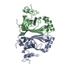
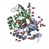
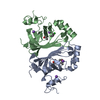
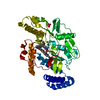
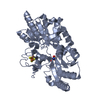
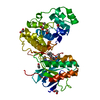
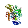
Crystal Structure of single point mutant Tyr20Phe p-coumaric Acid Decarboxylase from Lactobacillus plantarum: structural insights into the active site and decarboxylation catalytic mechanism
ComponentsP-COUMARIC ACID DECARBOXYLASE
KeywordsLYASE / ACTIVE SITE / COUMARIC ACIDS / DECARBOXYLATION / CATALYTIC MECHANISM
Function / homology
Function and homology information


Lyases; Carbon-carbon lyases; Carboxy-lyases / carboxy-lyase activity
Similarity search - Function
Phenolic acid decarboxylase / Phenolic acid decarboxylase (PAD) / Calycin beta-barrel core domain / Calycin / Lipocalin / Beta Barrel / Mainly Beta
Similarity search - Domain/homology
ACETATE ION / ISOPROPYL ALCOHOL / Phenolic acid decarboxylase / :
Similarity search - Component
Biological speciesLACTOBACILLUS PLANTARUM (bacteria)
MethodX-RAY DIFFRACTION / SYNCHROTRON / MOLECULAR REPLACEMENT / Resolution: 1.4 Å
AuthorsRodriguez, H. / Angulo, I. / de las Rivas, B. / Campillo, N. / Paez, J.A. / Munoz, R. / Mancheno, J.M.
CitationJournal: Proteins / Year: 2010
Title: P-Coumaric Acid Decarboxylase from Lactobacillus Plantarum: Structural Insights Into the Active Site and Decarboxylation Catalytic Mechanism.
Authors: Rodriguez, H. / Angulo, I. / D Elas Rivas, B. / Campillo, N. / Paez, J.A. / Munoz, R. / Mancheno, J.M.
History
DepositionOct 27, 2008Deposition site: PDBE / Processing site: PDBE
Revision 1.0Nov 17, 2009Provider: repository / Type: Initial release
Revision 1.1Nov 9, 2011Group: Database references / Derived calculations / Version format compliance
Revision 1.2Dec 13, 2023Group: Data collection / Database references ...Data collection / Database references / Derived calculations / Other / Refinement description
Category: chem_comp_atom / chem_comp_bond ...chem_comp_atom / chem_comp_bond / database_2 / pdbx_database_status / pdbx_initial_refinement_model / struct_ncs_dom_lim / struct_site
Item: _database_2.pdbx_DOI / _database_2.pdbx_database_accession ..._database_2.pdbx_DOI / _database_2.pdbx_database_accession / _pdbx_database_status.status_code_sf / _struct_ncs_dom_lim.beg_auth_comp_id / _struct_ncs_dom_lim.beg_label_asym_id / _struct_ncs_dom_lim.beg_label_comp_id / _struct_ncs_dom_lim.beg_label_seq_id / _struct_ncs_dom_lim.end_auth_comp_id / _struct_ncs_dom_lim.end_label_asym_id / _struct_ncs_dom_lim.end_label_comp_id / _struct_ncs_dom_lim.end_label_seq_id / _struct_site.pdbx_auth_asym_id / _struct_site.pdbx_auth_comp_id / _struct_site.pdbx_auth_seq_id
Remark 650 HELIX DETERMINATION METHOD: AUTHOR PROVIDED.
Remark 700 SHEET DETERMINATION METHOD: AUTHOR PROVIDED.

-
Structure visualization

Structure viewerMolecule:
MolmilJmol/JSmol

Downloads & links

-
Assembly

Deposited unit
A: P-COUMARIC ACID DECARBOXYLASE
B: P-COUMARIC ACID DECARBOXYLASE
hetero molecules


Theoretical massNumber of molelcules
Total (without water)45,8936
Polymers45,6552
Non-polymers2384
Water8,971498
1


  • Idetical with deposited unit
  • defined by author&software
TypeNameSymmetry operationNumber
identity operation1_555x,y,z1
Buried area3360 Å2
ΔGint-0.4 kcal/mol
Surface area14840 Å2
MethodPISA
Unit cell
Length a, b, c (Å)43.384, 43.384, 232.858
Angle α, β, γ (deg.)90.00, 90.00, 90.00
Int Tables number78
Space group name H-MP43
Noncrystallographic symmetry (NCS)NCS domain:
IDEns-IDDetails
11A
21B

NCS domain segments:

Component-ID: 1 / Ens-ID: 1 / Beg auth comp-ID: THR / Beg label comp-ID: THR / End auth comp-ID: LYS / End label comp-ID: LYS / Refine code: 5 / Auth seq-ID: 2 - 178 / Label seq-ID: 18 - 194

Dom-IDAuth asym-IDLabel asym-ID
1AA
2BB

-
Components

#1: Protein P-COUMARIC ACID DECARBOXYLASE


Mass: 22827.301 Da / Num. of mol.: 2 / Mutation: YES
Source method: isolated from a genetically manipulated source
Source: (gene. exp.) LACTOBACILLUS PLANTARUM (bacteria) / Production host: ESCHERICHIA COLI (E. coli) / Strain (production host): JM109(DE3) / References: UniProt: Q88RY7, UniProt: F9ULL2*PLUS
#2: Chemical ChemComp-ACT / ACETATE ION / Acetate


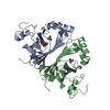
Mass: 59.044 Da / Num. of mol.: 2 / Source method: obtained synthetically / Formula: C2H3O2
#3: Chemical ChemComp-IPA / ISOPROPYL ALCOHOL / 2-PROPANOL / Isopropyl alcohol


Mass: 60.095 Da / Num. of mol.: 2 / Source method: obtained synthetically / Formula: C3H8O / Comment: alkaloid*YM
#4: Water ChemComp-HOH / water / Water


Mass: 18.015 Da / Num. of mol.: 498 / Source method: isolated from a natural source / Formula: H2O
Compound detailsENGINEERED RESIDUE IN CHAIN A, TYR 20 TO PHE ENGINEERED RESIDUE IN CHAIN B, TYR 20 TO PHE
Sequence detailsSINGLE POINT MUTANT (TYR20PHE) OF LACTOBACILLUS PLANTARUM P-COUMARIC ACID DECARBOXYLASE

-
Experimental details

-
Experiment

ExperimentMethod: X-RAY DIFFRACTION

-
Sample preparation

CrystalDensity Matthews: 2.6 Å3/Da / Density % sol: 52 % / Description: NONE

-
Data collection

DiffractionMean temperature: 100 K
Diffraction sourceSource: SYNCHROTRON / Site: ESRF / Beamline: ID29 / Wavelength: 0.9789
DetectorType: ADSC CCD / Detector: CCD / Date: Mar 5, 2008
RadiationProtocol: SINGLE WAVELENGTH / Monochromatic (M) / Laue (L): M / Scattering type: x-ray
Radiation wavelengthWavelength: 0.9789 Å / Relative weight: 1
ReflectionResolution: 1.4→58.22 Å / Num. obs: 76941 / % possible obs: 91.5 % / Redundancy: 6.7 % / Rmerge(I) obs: 0.09 / Net I/σ(I): 15.6
Reflection shellResolution: 1.4→1.48 Å / Redundancy: 2.8 % / Rmerge(I) obs: 0.39 / Mean I/σ(I) obs: 1.6 / % possible all: 91.5

-
Processing

Software
NameVersionClassification
REFMAC5.2.0019refinement
MOSFLMdata reduction
SCALAdata scaling
MOLREPphasing
RefinementMethod to determine structure: MOLECULAR REPLACEMENT
Starting model: PDB ENTRY 2W2A
Resolution: 1.4→58.22 Å / Cor.coef. Fo:Fc: 0.952 / Cor.coef. Fo:Fc free: 0.938 / SU B: 1.99 / SU ML: 0.042 / Cross valid method: THROUGHOUT / ESU R: 0.071 / ESU R Free: 0.071 / Stereochemistry target values: MAXIMUM LIKELIHOOD / Details: HYDROGENS HAVE BEEN ADDED IN THE RIDING POSITIONS.
RfactorNum. reflection% reflectionSelection details
Rfree0.221 3875 5 %RANDOM
Rwork0.199 ---
obs0.2 72949 91.5 %-
Solvent computationIon probe radii: 0.8 Å / Shrinkage radii: 0.8 Å / VDW probe radii: 1.2 Å / Solvent model: MASK
Displacement parametersBiso mean: 13.35 Å2
Baniso -1Baniso -2Baniso -3
1-0 Å20 Å20 Å2
2--0 Å20 Å2
3---0 Å2
Refinement stepCycle: LAST / Resolution: 1.4→58.22 Å
ProteinNucleic acidLigandSolventTotal
Num. atoms2950 0 16 498 3464
Refine LS restraints
Refine-IDTypeDev idealDev ideal targetNumber
X-RAY DIFFRACTIONr_bond_refined_d0.0060.0223052
X-RAY DIFFRACTIONr_bond_other_d
X-RAY DIFFRACTIONr_angle_refined_deg1.0721.9254142
X-RAY DIFFRACTIONr_angle_other_deg
X-RAY DIFFRACTIONr_dihedral_angle_1_deg5.9995352
X-RAY DIFFRACTIONr_dihedral_angle_2_deg34.51124.819166
X-RAY DIFFRACTIONr_dihedral_angle_3_deg12.85915500
X-RAY DIFFRACTIONr_dihedral_angle_4_deg14.3271510
X-RAY DIFFRACTIONr_chiral_restr0.0760.2422
X-RAY DIFFRACTIONr_gen_planes_refined0.0040.022392
X-RAY DIFFRACTIONr_gen_planes_other
X-RAY DIFFRACTIONr_nbd_refined0.1830.21448
X-RAY DIFFRACTIONr_nbd_other
X-RAY DIFFRACTIONr_nbtor_refined0.3110.22032
X-RAY DIFFRACTIONr_nbtor_other
X-RAY DIFFRACTIONr_xyhbond_nbd_refined0.0810.2449
X-RAY DIFFRACTIONr_xyhbond_nbd_other
X-RAY DIFFRACTIONr_metal_ion_refined
X-RAY DIFFRACTIONr_metal_ion_other
X-RAY DIFFRACTIONr_symmetry_vdw_refined0.1160.230
X-RAY DIFFRACTIONr_symmetry_vdw_other
X-RAY DIFFRACTIONr_symmetry_hbond_refined0.1740.247
X-RAY DIFFRACTIONr_symmetry_hbond_other
X-RAY DIFFRACTIONr_symmetry_metal_ion_refined
X-RAY DIFFRACTIONr_symmetry_metal_ion_other
X-RAY DIFFRACTIONr_mcbond_it0.5191.51823
X-RAY DIFFRACTIONr_mcbond_other
X-RAY DIFFRACTIONr_mcangle_it0.81222870
X-RAY DIFFRACTIONr_mcangle_other
X-RAY DIFFRACTIONr_scbond_it1.01331445
X-RAY DIFFRACTIONr_scbond_other
X-RAY DIFFRACTIONr_scangle_it1.6014.51272
X-RAY DIFFRACTIONr_scangle_other
X-RAY DIFFRACTIONr_long_range_B_refined
X-RAY DIFFRACTIONr_long_range_B_other
X-RAY DIFFRACTIONr_rigid_bond_restr
X-RAY DIFFRACTIONr_sphericity_free
X-RAY DIFFRACTIONr_sphericity_bonded
Refine LS restraints NCS

Ens-ID: 1 / Refine-ID: X-RAY DIFFRACTION

Dom-IDAuth asym-IDNumberTypeRms dev position (Å)Weight position
1A708medium positional0.030.5
2B708medium positional0.030.5
1A767loose positional0.155
2B767loose positional0.155
1A708medium thermal0.082
2B708medium thermal0.082
1A767loose thermal0.2110
2B767loose thermal0.2110
LS refinement shellResolution: 1.4→1.44 Å / Total num. of bins used: 20 /
RfactorNum. reflection
Rfree0.309 153
Rwork0.297 2846

+
About Yorodumi

-
News

-
Feb 9, 2022. New format data for meta-information of EMDB entries

New format data for meta-information of EMDB entries

  • Version 3 of the EMDB header file is now the official format.
  • The previous official version 1.9 will be removed from the archive.

Related info.:EMDB header

External links:wwPDB to switch to version 3 of the EMDB data model

-
Aug 12, 2020. Covid-19 info

Covid-19 info

URL: https://pdbj.org/emnavi/covid19.php

New page: Covid-19 featured information page in EM Navigator.

Related info.:Covid-19 info / Mar 5, 2020. Novel coronavirus structure data

+
Mar 5, 2020. Novel coronavirus structure data

Novel coronavirus structure data

Related info.:Yorodumi Speices / Aug 12, 2020. Covid-19 info

External links:COVID-19 featured content - PDBj / Molecule of the Month (242):Coronavirus Proteases

+
Jan 31, 2019. EMDB accession codes are about to change! (news from PDBe EMDB page)

EMDB accession codes are about to change! (news from PDBe EMDB page)

  • The allocation of 4 digits for EMDB accession codes will soon come to an end. Whilst these codes will remain in use, new EMDB accession codes will include an additional digit and will expand incrementally as the available range of codes is exhausted. The current 4-digit format prefixed with “EMD-” (i.e. EMD-XXXX) will advance to a 5-digit format (i.e. EMD-XXXXX), and so on. It is currently estimated that the 4-digit codes will be depleted around Spring 2019, at which point the 5-digit format will come into force.
  • The EM Navigator/Yorodumi systems omit the EMD- prefix.

Related info.:Q: What is EMD? / ID/Accession-code notation in Yorodumi/EM Navigator

External links:EMDB Accession Codes are Changing Soon! / Contact to PDBj

+
Jul 12, 2017. Major update of PDB

Major update of PDB

  • wwPDB released updated PDB data conforming to the new PDBx/mmCIF dictionary.
  • This is a major update changing the version number from 4 to 5, and with Remediation, in which all the entries are updated.
  • In this update, many items about electron microscopy experimental information are reorganized (e.g. em_software).
  • Now, EM Navigator and Yorodumi are based on the updated data.

External links:wwPDB Remediation / Enriched Model Files Conforming to OneDep Data Standards Now Available in the PDB FTP Archive

-
Yorodumi

Thousand views of thousand structures

  • Yorodumi is a browser for structure data from EMDB, PDB, SASBDB, etc.
  • This page is also the successor to EM Navigator detail page, and also detail information page/front-end page for Omokage search.
  • The word "yorodu" (or yorozu) is an old Japanese word meaning "ten thousand". "mi" (miru) is to see.

Related info.:EMDB / PDB / SASBDB / Comparison of 3 databanks / Yorodumi Search / Aug 31, 2016. New EM Navigator & Yorodumi / Yorodumi Papers / Jmol/JSmol / Function and homology information / Changes in new EM Navigator and Yorodumi

Read more