 Movie
Movie Controller

[English] 日本語
 Yorodumi
- PDB-2vyr: Structure of human MDM4 N-terminal domain bound to a single domai... -
+ Open data

- Basic information
| Entry | Database: PDB / ID: 2vyr | ||||||
|---|---|---|---|---|---|---|---|
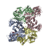
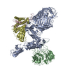
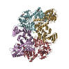
| Title | Structure of human MDM4 N-terminal domain bound to a single domain antibody | ||||||
 Components Components |
| ||||||
 Keywords Keywords | IMMUNE SYSTEM / NUCLEUS / HUMAN MDM4 / ZINC-FINGER | ||||||
| Function / homology |  Function and homology information Function and homology informationatrial septum development / heart valve development / atrioventricular valve morphogenesis / endocardial cushion morphogenesis / ventricular septum development / negative regulation of signal transduction by p53 class mediator / transcription repressor complex / DNA damage response, signal transduction by p53 class mediator / Stabilization of p53 / negative regulation of protein catabolic process ...atrial septum development / heart valve development / atrioventricular valve morphogenesis / endocardial cushion morphogenesis / ventricular septum development / negative regulation of signal transduction by p53 class mediator / transcription repressor complex / DNA damage response, signal transduction by p53 class mediator / Stabilization of p53 / negative regulation of protein catabolic process / Oncogene Induced Senescence / Regulation of TP53 Activity through Methylation / enzyme activator activity / ubiquitin-protein transferase activity / Regulation of TP53 Degradation / protein-containing complex assembly / Oxidative Stress Induced Senescence / cellular response to hypoxia / Regulation of TP53 Activity through Phosphorylation / regulation of cell cycle / protein stabilization / Ub-specific processing proteases / protein ubiquitination / negative regulation of cell population proliferation / negative regulation of DNA-templated transcription / negative regulation of apoptotic process / enzyme binding / negative regulation of transcription by RNA polymerase II / zinc ion binding / nucleoplasm / nucleus Similarity search - Function | ||||||
| Biological species |  HOMO SAPIENS (human) HOMO SAPIENS (human) | ||||||
| Method |  X-RAY DIFFRACTION / SYNCHROTRON / MAD / Resolution: 2 Å X-RAY DIFFRACTION / SYNCHROTRON / MAD / Resolution: 2 Å | ||||||
 Authors Authors | Yu, G.W. / Vaysburd, M. / Allen, M.D. / Settanni, G. / Fersht, A.R. | ||||||
 Citation Citation | Journal: J.Mol.Biol. / Year: 2009 Title: Structure of Human Mdm4 N-Terminal Domain Bound to a Single-Domain Antibody. Authors: Yu, G.W. / Vaysburd, M. / Allen, M.D. / Settanni, G. / Fersht, A.R. | ||||||
| History |
| ||||||
| Remark 700 | SHEET THE SHEET STRUCTURE OF THIS MOLECULE IS BIFURCATED. IN ORDER TO REPRESENT THIS FEATURE IN ... SHEET THE SHEET STRUCTURE OF THIS MOLECULE IS BIFURCATED. IN ORDER TO REPRESENT THIS FEATURE IN THE SHEET RECORDS BELOW, TWO SHEETS ARE DEFINED. |
- Structure visualization
| Structure viewer | Molecule: MolmilJmol/JSmol |
|---|
- Downloads & links
-Download
| PDBx/mmCIF format | 2vyr.cif.gz | 283.4 KB | Display | PDBx/mmCIF format |
|---|---|---|---|---|
| PDB format | pdb2vyr.ent.gz | 229.3 KB | Display | PDB format |
| PDBx/mmJSON format | 2vyr.json.gz | Tree view | PDBx/mmJSON format | |
| Others |  Other downloads Other downloads |
-Validation report
| Arichive directory | https://data.pdbj.org/pub/pdb/validation_reports/vy/2vyrftp://data.pdbj.org/pub/pdb/validation_reports/vy/2vyr | HTTPS FTP |
|---|
-Related structure data
| Related structure data | |
|---|---|
| Similar structure data |


-Links
 PDBj
PDBj


- Assembly
| Deposited unit | 
| ||||||||
|---|---|---|---|---|---|---|---|---|---|
| 1 |
| ||||||||
| Unit cell |
|
-Components
| #1: Protein | Mass: 11306.168 Da / Num. of mol.: 4 / Fragment: RESIDUES 16-116 Source method: isolated from a genetically manipulated source Source: (gene. exp.) HOMO SAPIENS (human) / Production host:  #2: Antibody | Mass: 17041.904 Da / Num. of mol.: 8 Source method: isolated from a genetically manipulated source Source: (gene. exp.) HOMO SAPIENS (human) / Description: NON-BIOLOGICAL SEQUENCE, OBTAINED BY SELECTION / Production host: #3: Chemical | ChemComp-SO4 /   Mass: 96.063 Da / Num. of mol.: 4 / Source method: obtained synthetically / Formula: SO4 Mass: 96.063 Da / Num. of mol.: 4 / Source method: obtained synthetically / Formula: SO4#4: Water | ChemComp-HOH / |  Mass: 18.015 Da / Num. of mol.: 752 / Source method: isolated from a natural source / Formula: H2O Mass: 18.015 Da / Num. of mol.: 752 / Source method: isolated from a natural source / Formula: H2OHas protein modification | Y | |
|---|
-Experimental details
-Experiment
| Experiment | Method: X-RAY DIFFRACTION / Number of used crystals: 2 |
|---|
- Sample preparation
| Crystal | Density Matthews: 2.44 Å3/Da / Density % sol: 50 % Description: STRUCTURE WAS INITIALLY SOLVED USING MAD ON A SE- MET COMPLEX CONTAINING LABELLED MDMX. THE NATIVE STRUCTURE WAS SUBSEQUENTLY DETERMINED BY MOLECULAR REPLACEMENT |
|---|---|
| Crystal grow | pH: 7 Details: SE-MET: 20% PEG 3350, 0.2 M MGCL2, 1MM TRIS PH 7.0, 5MM B-ME, PROTEIN 10MG/ML. NATIVE: 1.6M AMMONIUM SULPHATE, 0.5M LITHIUM CHLORIDE, 1MM TRIS, PH 7.0, 5MM B-ME, PROTEIN 10MG/ML |
-Data collection
| Diffraction | Mean temperature: 100 K |
|---|---|
| Diffraction source | Source: SYNCHROTRON / Site: ESRF  / Beamline: ID14-1 / Wavelength: 0.954 / Beamline: ID14-1 / Wavelength: 0.954 |
| Detector | Type: ADSC CCD / Detector: CCD |
| Radiation | Protocol: MAD / Monochromatic (M) / Laue (L): M / Scattering type: x-ray |
| Radiation wavelength | Wavelength: 0.954 Å / Relative weight: 1 |
| Reflection | Resolution: 2→30 Å / Num. obs: 116657 / % possible obs: 99.6 % / Observed criterion σ(I): 2 / Redundancy: 3.9 % / Rmerge(I) obs: 0.09 / Net I/σ(I): 10.4 |
| Reflection shell | Highest resolution: 2 Å / Redundancy: 4 % / Rmerge(I) obs: 0.27 / Mean I/σ(I) obs: 4.3 / % possible all: 99.6 |
- Processing
| Software |
| |||||||||||||||||||||||||||||||||||||||||||||||||||||||||||||||||||||||||||||||||||||||||||||||||||||||||||||||||||||||||||||||||||||||||||||||||||||||||||||||||||||||||||||||||||||||||||||||||||||||||||||||||||||||||
|---|---|---|---|---|---|---|---|---|---|---|---|---|---|---|---|---|---|---|---|---|---|---|---|---|---|---|---|---|---|---|---|---|---|---|---|---|---|---|---|---|---|---|---|---|---|---|---|---|---|---|---|---|---|---|---|---|---|---|---|---|---|---|---|---|---|---|---|---|---|---|---|---|---|---|---|---|---|---|---|---|---|---|---|---|---|---|---|---|---|---|---|---|---|---|---|---|---|---|---|---|---|---|---|---|---|---|---|---|---|---|---|---|---|---|---|---|---|---|---|---|---|---|---|---|---|---|---|---|---|---|---|---|---|---|---|---|---|---|---|---|---|---|---|---|---|---|---|---|---|---|---|---|---|---|---|---|---|---|---|---|---|---|---|---|---|---|---|---|---|---|---|---|---|---|---|---|---|---|---|---|---|---|---|---|---|---|---|---|---|---|---|---|---|---|---|---|---|---|---|---|---|---|---|---|---|---|---|---|---|---|---|---|---|---|---|---|---|---|
| Refinement | Method to determine structure: MAD Starting model: NONE Resolution: 2→25 Å / SU ML: 0.32 / Phase error: 23.59 / Stereochemistry target values: ML / Details: DISORDERED REGIONS WERE MODELED STEREOCHEMICALLY
| |||||||||||||||||||||||||||||||||||||||||||||||||||||||||||||||||||||||||||||||||||||||||||||||||||||||||||||||||||||||||||||||||||||||||||||||||||||||||||||||||||||||||||||||||||||||||||||||||||||||||||||||||||||||||
| Solvent computation | Shrinkage radii: 0.9 Å / VDW probe radii: 1.11 Å / Solvent model: FLAT BULK SOLVENT MODEL / Bsol: 46.385 Å2 / ksol: 0.346 e/Å3 | |||||||||||||||||||||||||||||||||||||||||||||||||||||||||||||||||||||||||||||||||||||||||||||||||||||||||||||||||||||||||||||||||||||||||||||||||||||||||||||||||||||||||||||||||||||||||||||||||||||||||||||||||||||||||
| Refinement step | Cycle: LAST / Resolution: 2→25 Å
| |||||||||||||||||||||||||||||||||||||||||||||||||||||||||||||||||||||||||||||||||||||||||||||||||||||||||||||||||||||||||||||||||||||||||||||||||||||||||||||||||||||||||||||||||||||||||||||||||||||||||||||||||||||||||
| Refine LS restraints |
| |||||||||||||||||||||||||||||||||||||||||||||||||||||||||||||||||||||||||||||||||||||||||||||||||||||||||||||||||||||||||||||||||||||||||||||||||||||||||||||||||||||||||||||||||||||||||||||||||||||||||||||||||||||||||
| LS refinement shell |
|
