 Movie
Movie Controller

[English] 日本語
 Yorodumi
- PDB-2vw9: Single stranded DNA binding protein complex from Helicobacter pylori -
+ Open data

- Basic information
| Entry | Database: PDB / ID: 2vw9 | ||||||
|---|---|---|---|---|---|---|---|
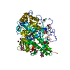
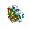
| Title | Single stranded DNA binding protein complex from Helicobacter pylori | ||||||
 Components Components |
| ||||||
 Keywords Keywords | DNA BINDING PROTEIN / DNA REPLICATION / SINGLE-STRANDED DNA / SINGLE-STRANDED DNA BINDING PROTEIN / OLIGONUCLEOTIDE/OLIGOSACCHARIDE BINDING FOLD / HELICOBACTER PYLORI / OB-FOLD / DNA DAMAGE / DNA REPAIR / DNA-BINDING PROTEIN | ||||||
| Function / homology |  Function and homology information Function and homology informationnucleoid / enzyme activator activity / single-stranded DNA binding / DNA recombination / DNA replication / DNA repair Similarity search - Function | ||||||
| Biological species |   HELICOBACTER PYLORI (bacteria) HELICOBACTER PYLORI (bacteria) | ||||||
| Method |  X-RAY DIFFRACTION / SYNCHROTRON / MOLECULAR REPLACEMENT / Resolution: 2.3 Å X-RAY DIFFRACTION / SYNCHROTRON / MOLECULAR REPLACEMENT / Resolution: 2.3 Å | ||||||
 Authors Authors | Chan, K.-W. / Wang, C.-H. / Lee, Y.-J. / Sun, Y.-J. | ||||||
 Citation Citation | Journal: J.Mol.Biol. / Year: 2009 Title: Single-Stranded DNA-Binding Protein Complex from Helicobacter Pylori Suggests an Ssdna-Binding Surface. Authors: Chan, K.-W. / Lee, Y.-J. / Wang, C.-H. / Huang, H. / Sun, Y.-J. | ||||||
| History |
| ||||||
| Remark 700 | SHEET DETERMINATION METHOD: DSSP THE SHEETS PRESENTED AS "AA", "BA" IN EACH CHAIN ON SHEET RECORDS ... SHEET DETERMINATION METHOD: DSSP THE SHEETS PRESENTED AS "AA", "BA" IN EACH CHAIN ON SHEET RECORDS BELOW IS ACTUALLY AN 7-STRANDED BARREL THIS IS REPRESENTED BY A 8-STRANDED SHEET IN WHICH THE FIRST AND LAST STRANDS ARE IDENTICAL. |
- Structure visualization
| Structure viewer | Molecule: MolmilJmol/JSmol |
|---|
- Downloads & links
-Download
| PDBx/mmCIF format | 2vw9.cif.gz | 71 KB | Display | PDBx/mmCIF format |
|---|---|---|---|---|
| PDB format | pdb2vw9.ent.gz | 51.1 KB | Display | PDB format |
| PDBx/mmJSON format | 2vw9.json.gz | Tree view | PDBx/mmJSON format | |
| Others |  Other downloads Other downloads |
-Validation report
| Arichive directory | https://data.pdbj.org/pub/pdb/validation_reports/vw/2vw9ftp://data.pdbj.org/pub/pdb/validation_reports/vw/2vw9 | HTTPS FTP |
|---|
-Related structure data
| Related structure data |  1eygS S: Starting model for refinement |
|---|---|
| Similar structure data |


-Links
 PDBj
PDBj


- Assembly
| Deposited unit | 
| ||||||||
|---|---|---|---|---|---|---|---|---|---|
| 1 | 
| ||||||||
| Unit cell |
|
-Components
| #1: Protein | Mass: 14968.873 Da / Num. of mol.: 2 / Fragment: RESIDUES 1-134 Source method: isolated from a genetically manipulated source Source: (gene. exp.) HELICOBACTER PYLORI (bacteria) / Strain: 26695 / Plasmid: PQE30 / Production host: #2: DNA chain | | Mass: 10601.791 Da / Num. of mol.: 1 / Source method: obtained synthetically #3: Water | ChemComp-HOH / |  Mass: 18.015 Da / Num. of mol.: 160 / Source method: isolated from a natural source / Formula: H2O Mass: 18.015 Da / Num. of mol.: 160 / Source method: isolated from a natural source / Formula: H2O |
|---|
-Experimental details
-Experiment
| Experiment | Method: X-RAY DIFFRACTION |
|---|
- Sample preparation
| Crystal | Density Matthews: 2.25 Å3/Da / Density % sol: 45 % / Description: NONE |
|---|---|
| Crystal grow | Details: SODIUM CITRATE BUFFER PH5.5, 1.0 M LI2SO4 AND 0.5 M (NH4)2SO4 |
-Data collection
| Diffraction | Mean temperature: 100 K |
|---|---|
| Diffraction source | Source: SYNCHROTRON / Site: SPring-8  / Beamline: BL12B2 / Wavelength: 1 / Beamline: BL12B2 / Wavelength: 1 |
| Detector | Type: ADSC CCD / Detector: CCD / Date: Mar 15, 2007 |
| Radiation | Protocol: SINGLE WAVELENGTH / Monochromatic (M) / Laue (L): M / Scattering type: x-ray |
| Radiation wavelength | Wavelength: 1 Å / Relative weight: 1 |
| Reflection | Resolution: 2.3→30 Å / Num. obs: 17468 / % possible obs: 98 % / Observed criterion σ(I): 2 / Redundancy: 7 % / Biso Wilson estimate: 24.6 Å2 / Rmerge(I) obs: 0.05 / Net I/σ(I): 51.7 |
| Reflection shell | Resolution: 2.3→2.38 Å / Redundancy: 7.1 % / Rmerge(I) obs: 0.47 / Mean I/σ(I) obs: 6.2 / % possible all: 100 |
- Processing
| Software |
| ||||||||||||||||||||||||||||||||||||||||||||||||||||||||||||
|---|---|---|---|---|---|---|---|---|---|---|---|---|---|---|---|---|---|---|---|---|---|---|---|---|---|---|---|---|---|---|---|---|---|---|---|---|---|---|---|---|---|---|---|---|---|---|---|---|---|---|---|---|---|---|---|---|---|---|---|---|---|
| Refinement | Method to determine structure: MOLECULAR REPLACEMENT Starting model: PDB ENTRY 1EYG Resolution: 2.3→25.13 Å / Rfactor Rfree error: 0.009 / Data cutoff high absF: 42178.93 / Isotropic thermal model: RESTRAINED / Cross valid method: THROUGHOUT / σ(F): 2 Details: BULK SOLVENT MODEL USED MISSING REGIONS CHAIN A RESIDUES 1109-1134 CHAIN B RESIDUES 2106-2134 SSDNA 6, 10-13, 17, 27-29, 34-35 FOLLOW RESIDUES ARE REPLACED TO ALA CHAIN A K1038, K1039, K1108 ...Details: BULK SOLVENT MODEL USED MISSING REGIONS CHAIN A RESIDUES 1109-1134 CHAIN B RESIDUES 2106-2134 SSDNA 6, 10-13, 17, 27-29, 34-35 FOLLOW RESIDUES ARE REPLACED TO ALA CHAIN A K1038, K1039, K1108 CHAIN B K2038, K2039, D2041
| ||||||||||||||||||||||||||||||||||||||||||||||||||||||||||||
| Solvent computation | Solvent model: FLAT MODEL / Bsol: 61.1119 Å2 / ksol: 0.4 e/Å3 | ||||||||||||||||||||||||||||||||||||||||||||||||||||||||||||
| Displacement parameters | Biso mean: 49.8 Å2
| ||||||||||||||||||||||||||||||||||||||||||||||||||||||||||||
| Refine analyze |
| ||||||||||||||||||||||||||||||||||||||||||||||||||||||||||||
| Refinement step | Cycle: LAST / Resolution: 2.3→25.13 Å
| ||||||||||||||||||||||||||||||||||||||||||||||||||||||||||||
| Refine LS restraints |
| ||||||||||||||||||||||||||||||||||||||||||||||||||||||||||||
| Refine LS restraints NCS | NCS model details: NONE | ||||||||||||||||||||||||||||||||||||||||||||||||||||||||||||
| LS refinement shell | Resolution: 2.3→2.44 Å / Rfactor Rfree error: 0.022 / Total num. of bins used: 6
| ||||||||||||||||||||||||||||||||||||||||||||||||||||||||||||
| Xplor file | Serial no: 1 / Param file: PROTEIN_REP.PARAM / Topol file: WATER_REP.PARAM |
