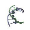
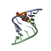
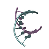
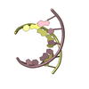
- PDB-2vuq: Crystal structure of a human tRNAGly acceptor stem microhelix (de... -
+
Open data
ID or keywords:
Loading...
-
Basic information
Entry
Database: PDB / ID: 2vuq
Title
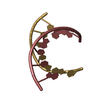
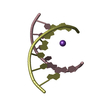
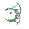
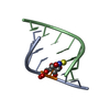
Crystal structure of a human tRNAGly acceptor stem microhelix (derived from the gene sequence DG9990) at 1.18 Angstroem resolution
Components
5'-R(*CP*CP*AP*AP*UP*GP*CP)-3'
5'-R(*GP*CP*AP*UP*UP*GP*GP)-3'
Keywords
RNA / CLASS II AMINOACYL-TRNA / SYNTHETASE SYSTEM / TRNA IDENTITY ELEMENTS / GLYCYL-TRNA-SYNTHETASE (GLYRS) / ISOACCEPTORS / HUMAN TRNAGLY / AMINOACYL-TRNA-STEM
Mass: 18.015 Da / Num. of mol.: 48 / Source method: isolated from a natural source / Formula: H2O
-
Experimental details
-
Experiment
Experiment
Method: X-RAY DIFFRACTION / Number of used crystals: 1
-
Sample preparation
Crystal
Density Matthews: 2.07 Å3/Da / Density % sol: 40.21 % / Description: NONE
Crystal grow
Temperature: 294 K / pH: 6 Details: 10% (V/V) MPD, 40 MM SODIUM CACODYLATE, PH 6.0, 12 MM SPERMINE TETRACHLORIDE, 80 MM POTASSIUM CHLORIDE, EQUILIBRATED AGAINST 35 % (V/V) MPD AT 294 K.
Resolution: 1.18→22.25 Å / Cor.coef. Fo:Fc: 0.929 / Cor.coef. Fo:Fc free: 0.93 / SU B: 1.048 / SU ML: 0.023 / Cross valid method: THROUGHOUT / ESU R: 0.05 / ESU R Free: 0.048 / Stereochemistry target values: MAXIMUM LIKELIHOOD / Details: HYDROGENS HAVE BEEN ADDED IN THE RIDING POSITIONS.
 Movie
Movie Controller

 Yorodumi
 Open data
 Basic information
 Components
 Keywords
 Function and homology information
 HOMO SAPIENS (human)
 X-RAY DIFFRACTION /
  Authors
 Citation
 Structure visualization
 Downloads & links
 Other downloads
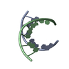
 PDBj
PDBj Assembly
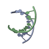
 Mass: 18.015 Da / Num. of mol.: 48 / Source method: isolated from a natural source / Formula: H2O
 Sample preparation
 / Beamline: I04 / Wavelength: 0.9696
  Processing