 Movie
Movie Controller

[English] 日本語
 Yorodumi
- PDB-2vma: The three-dimensional structure of the cytoplasmic domains of Eps... -
+ Open data

- Basic information
| Entry | Database: PDB / ID: 2vma | ||||||
|---|---|---|---|---|---|---|---|
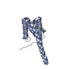
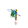
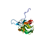
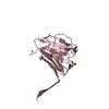
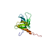
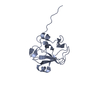
| Title | The three-dimensional structure of the cytoplasmic domains of EpsF from the Type 2 Secretion System of Vibrio cholerae | ||||||
 Components Components | GENERAL SECRETION PATHWAY PROTEIN F | ||||||
 Keywords Keywords | TRANSPORT PROTEIN / TRANSMEMBRANE / INNER MEMBRANE / TYPE 2 SECRETION / T4PB / T2SS / VIBRIO / CHOLERA / MEMBRANE / TRANSPORT / PROTEIN SECRETION | ||||||
| Function / homology |  Function and homology information Function and homology informationprotein secretion by the type II secretion system / type II protein secretion system complex / metal ion binding / plasma membrane Similarity search - Function | ||||||
| Biological species |   VIBRIO CHOLERAE (bacteria) VIBRIO CHOLERAE (bacteria) | ||||||
| Method |  X-RAY DIFFRACTION / SAD / Resolution: 1.9 Å X-RAY DIFFRACTION / SAD / Resolution: 1.9 Å | ||||||
 Authors Authors | Abendroth, J. / Korotkov, K.V. / Mitchell, D.D. / Kreger, A. / Hol, W.G.J. | ||||||
 Citation Citation | Journal: J.Struct.Biol. / Year: 2009 Title: The Three-Dimensional Structure of the Cytoplasmic Domains of Epsf from the Type 2 Secretion System of Vibrio Cholerae. Authors: Abendroth, J. / Mitchell, D.D. / Korotkov, K.V. / Johnson, T.L. / Kreger, A. / Sandkvist, M. / Hol, W.G. | ||||||
| History |
|
- Structure visualization
| Structure viewer | Molecule: MolmilJmol/JSmol |
|---|
- Downloads & links
-Download
| PDBx/mmCIF format | 2vma.cif.gz | 66.4 KB | Display | PDBx/mmCIF format |
|---|---|---|---|---|
| PDB format | pdb2vma.ent.gz | 50.5 KB | Display | PDB format |
| PDBx/mmJSON format | 2vma.json.gz | Tree view | PDBx/mmJSON format | |
| Others |  Other downloads Other downloads |
-Validation report
| Arichive directory | https://data.pdbj.org/pub/pdb/validation_reports/vm/2vmaftp://data.pdbj.org/pub/pdb/validation_reports/vm/2vma | HTTPS FTP |
|---|
-Related structure data


-Links
 PDBj
PDBj
- Assembly
| Deposited unit | 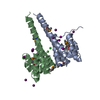
| |||||||||||||||||||||||||||
|---|---|---|---|---|---|---|---|---|---|---|---|---|---|---|---|---|---|---|---|---|---|---|---|---|---|---|---|---|
| 1 | 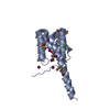
| |||||||||||||||||||||||||||
| 2 | 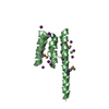
| |||||||||||||||||||||||||||
| Unit cell |
| |||||||||||||||||||||||||||
| Noncrystallographic symmetry (NCS) | NCS domain:
NCS domain segments:
NCS oper: (Code: given Matrix: (-0.99988, 0.00497, -0.01461), Vector: |
-Components
| #1: Protein | Mass: 14028.238 Da / Num. of mol.: 2 / Fragment: RESIDUES 56-170 Source method: isolated from a genetically manipulated source Details: RESIDUES 56-170, PLUS C-TERMINAL TEV- CLEAVABLE HIS6-TAG Source: (gene. exp.) VIBRIO CHOLERAE (bacteria) / Plasmid: PACYC-CT / Production host: #2: Chemical |   Mass: 40.078 Da / Num. of mol.: 3 / Source method: obtained synthetically / Formula: Ca Mass: 40.078 Da / Num. of mol.: 3 / Source method: obtained synthetically / Formula: Ca#3: Chemical | ChemComp-IOD /   Mass: 126.904 Da / Num. of mol.: 15 / Source method: obtained synthetically / Formula: I Mass: 126.904 Da / Num. of mol.: 15 / Source method: obtained synthetically / Formula: I#4: Water | ChemComp-HOH / |  Mass: 18.015 Da / Num. of mol.: 215 / Source method: isolated from a natural source / Formula: H2O Mass: 18.015 Da / Num. of mol.: 215 / Source method: isolated from a natural source / Formula: H2OHas protein modification | Y | Sequence details | CYTOPLASMI | |
|---|
-Experimental details
-Experiment
| Experiment | Method: X-RAY DIFFRACTION / Number of used crystals: 1 |
|---|
- Sample preparation
| Crystal | Density Matthews: 2 Å3/Da / Density % sol: 38.08 % / Description: NONE |
|---|---|
| Crystal grow | pH: 7 Details: 1UL PROTEIN, 1UL RESERVOIR: 12.5% PEG 400, 200MM CAOAC2, 100MM MES PH 7.0 |
-Data collection
| Diffraction | Mean temperature: 100 K |
|---|---|
| Diffraction source | Source: ROTATING ANODE / Wavelength: 1.5418 |
| Detector | Type: RIGAKU CCD / Detector: CCD / Date: Oct 23, 2006 / Details: MIRRORS |
| Radiation | Monochromator: OSMIC VARIMAX / Protocol: SINGLE WAVELENGTH / Monochromatic (M) / Laue (L): M / Scattering type: x-ray |
| Radiation wavelength | Wavelength: 1.5418 Å / Relative weight: 1 |
| Reflection | Resolution: 1.9→48.82 Å / Num. obs: 18609 / % possible obs: 96.6 % / Observed criterion σ(I): 0 / Redundancy: 12.11 % / Rmerge(I) obs: 0.09 / Net I/σ(I): 16.1 |
| Reflection shell | Resolution: 1.9→1.97 Å / Redundancy: 3.9 % / Rmerge(I) obs: 0.33 / Mean I/σ(I) obs: 3.9 / % possible all: 77.2 |
- Processing
| Software |
| ||||||||||||||||||||||||||||||||||||||||||||||||||||||||||||||||||||||||||||||||||||||||||||||||||||||||||||||||||||||||||||||||||||||||||||||||||||||||||||||||||||||||||||||||||||||
|---|---|---|---|---|---|---|---|---|---|---|---|---|---|---|---|---|---|---|---|---|---|---|---|---|---|---|---|---|---|---|---|---|---|---|---|---|---|---|---|---|---|---|---|---|---|---|---|---|---|---|---|---|---|---|---|---|---|---|---|---|---|---|---|---|---|---|---|---|---|---|---|---|---|---|---|---|---|---|---|---|---|---|---|---|---|---|---|---|---|---|---|---|---|---|---|---|---|---|---|---|---|---|---|---|---|---|---|---|---|---|---|---|---|---|---|---|---|---|---|---|---|---|---|---|---|---|---|---|---|---|---|---|---|---|---|---|---|---|---|---|---|---|---|---|---|---|---|---|---|---|---|---|---|---|---|---|---|---|---|---|---|---|---|---|---|---|---|---|---|---|---|---|---|---|---|---|---|---|---|---|---|---|---|
| Refinement | Method to determine structure: SAD Starting model: NONE Resolution: 1.9→46.32 Å / Cor.coef. Fo:Fc: 0.946 / Cor.coef. Fo:Fc free: 0.933 / SU B: 6.078 / SU ML: 0.112 / TLS residual ADP flag: LIKELY RESIDUAL / Cross valid method: THROUGHOUT / ESU R: 0.194 / ESU R Free: 0.172 / Stereochemistry target values: MAXIMUM LIKELIHOOD / Details: HYDROGENS HAVE BEEN ADDED IN THE RIDING POSITIONS.
| ||||||||||||||||||||||||||||||||||||||||||||||||||||||||||||||||||||||||||||||||||||||||||||||||||||||||||||||||||||||||||||||||||||||||||||||||||||||||||||||||||||||||||||||||||||||
| Solvent computation | Ion probe radii: 0.8 Å / Shrinkage radii: 0.8 Å / VDW probe radii: 1.4 Å / Solvent model: MASK | ||||||||||||||||||||||||||||||||||||||||||||||||||||||||||||||||||||||||||||||||||||||||||||||||||||||||||||||||||||||||||||||||||||||||||||||||||||||||||||||||||||||||||||||||||||||
| Displacement parameters | Biso mean: 17.5 Å2
| ||||||||||||||||||||||||||||||||||||||||||||||||||||||||||||||||||||||||||||||||||||||||||||||||||||||||||||||||||||||||||||||||||||||||||||||||||||||||||||||||||||||||||||||||||||||
| Refinement step | Cycle: LAST / Resolution: 1.9→46.32 Å
| ||||||||||||||||||||||||||||||||||||||||||||||||||||||||||||||||||||||||||||||||||||||||||||||||||||||||||||||||||||||||||||||||||||||||||||||||||||||||||||||||||||||||||||||||||||||
| Refine LS restraints |
|