 Movie
Movie Controller

[English] 日本語
 Yorodumi
- PDB-2v0l: Characterization of Substrate Binding and Catalysis of the Potent... -
+ Open data

- Basic information
| Entry | Database: PDB / ID: 2v0l | ||||||
|---|---|---|---|---|---|---|---|
| Title | Characterization of Substrate Binding and Catalysis of the Potential Antibacterial Target N-acetylglucosamine-1-phosphate Uridyltransferase (GlmU) | ||||||
 Components Components | BIFUNCTIONAL PROTEIN GLMU | ||||||
 Keywords Keywords | TRANSFERASE / GLMU / CELL WALL / MAGNESIUM / CELL SHAPE / PEPTIDOGLYCAN SYNTHESIS / MULTIFUNCTIONAL ENZYME / NUCLEOTIDYLTRANSFERASE / URIDYLATION / METAL-BINDING / ACYLTRANSFERASE / CATALYTIC MECHANISM / ASSOCIATIVE MECHANISM | ||||||
| Function / homology |  Function and homology information Function and homology informationglucosamine-1-phosphate N-acetyltransferase / glucosamine-1-phosphate N-acetyltransferase activity / UDP-N-acetylglucosamine diphosphorylase / UDP-N-acetylglucosamine diphosphorylase activity / UDP-N-acetylglucosamine biosynthetic process / lipid A biosynthetic process / peptidoglycan biosynthetic process / cell wall organization / cell morphogenesis / regulation of cell shape ...glucosamine-1-phosphate N-acetyltransferase / glucosamine-1-phosphate N-acetyltransferase activity / UDP-N-acetylglucosamine diphosphorylase / UDP-N-acetylglucosamine diphosphorylase activity / UDP-N-acetylglucosamine biosynthetic process / lipid A biosynthetic process / peptidoglycan biosynthetic process / cell wall organization / cell morphogenesis / regulation of cell shape / magnesium ion binding / membrane / cytosol Similarity search - Function | ||||||
| Biological species |  HAEMOPHILUS INFLUENZAE (bacteria) HAEMOPHILUS INFLUENZAE (bacteria) | ||||||
| Method |  X-RAY DIFFRACTION / SYNCHROTRON / MOLECULAR REPLACEMENT / Resolution: 2.2 Å X-RAY DIFFRACTION / SYNCHROTRON / MOLECULAR REPLACEMENT / Resolution: 2.2 Å | ||||||
 Authors Authors | Mochalkin, I. / Lightle, S. / Ohren, J.F. / Chirgadze, N.Y. | ||||||
 Citation Citation | Journal: Protein Sci. / Year: 2007 Title: Characterization of Substrate Binding and Catalysis in the Potential Antibacterial Target N-Acetylglucosamine-1-Phosphate Uridyltransferase (Glmu). Authors: Mochalkin, I. / Lightle, S. / Zhu, Y. / Ohren, J.F. / Spessard, C. / Chirgadze, N.Y. / Banotai, C. / Melnick, M. / Mcdowell, L. | ||||||
| History |
| ||||||
| Remark 700 | SHEET THE SHEET STRUCTURE OF THIS MOLECULE IS BIFURCATED. IN ORDER TO REPRESENT THIS FEATURE IN ... SHEET THE SHEET STRUCTURE OF THIS MOLECULE IS BIFURCATED. IN ORDER TO REPRESENT THIS FEATURE IN THE SHEET RECORDS BELOW, TWO SHEETS ARE DEFINED. |
- Structure visualization
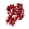
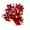
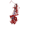
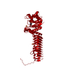
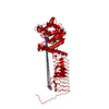
| Structure viewer | Molecule: MolmilJmol/JSmol |
|---|
- Downloads & links
-Download
| PDBx/mmCIF format | 2v0l.cif.gz | 110.6 KB | Display | PDBx/mmCIF format |
|---|---|---|---|---|
| PDB format | pdb2v0l.ent.gz | 84 KB | Display | PDB format |
| PDBx/mmJSON format | 2v0l.json.gz | Tree view | PDBx/mmJSON format | |
| Others |  Other downloads Other downloads |
-Validation report
| Arichive directory | https://data.pdbj.org/pub/pdb/validation_reports/v0/2v0lftp://data.pdbj.org/pub/pdb/validation_reports/v0/2v0l | HTTPS FTP |
|---|
-Related structure data
| Related structure data |  2v0hSC  2v0iC  2v0jC 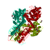 2v0kC S: Starting model for refinement C: citing same article ( |
|---|---|
| Similar structure data |


-Links
 PDBj
PDBj


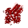
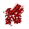
- Assembly
| Deposited unit | 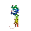
| ||||||||
|---|---|---|---|---|---|---|---|---|---|
| 1 | 
| ||||||||
| Unit cell |
|
-Components
-Protein , 1 types, 1 molecules A
| #1: Protein | Mass: 49346.062 Da / Num. of mol.: 1 / Source method: isolated from a natural source / Source: (natural) HAEMOPHILUS INFLUENZAE (bacteria) / References: UniProt: P43889, Transferases |
|---|
-Non-polymers , 5 types, 423 molecules 


| #2: Chemical | ChemComp-URI /  Mass: 244.201 Da / Num. of mol.: 1 / Source method: obtained synthetically / Formula: C9H12N2O6 Mass: 244.201 Da / Num. of mol.: 1 / Source method: obtained synthetically / Formula: C9H12N2O6 | ||||
|---|---|---|---|---|---|
| #3: Chemical | ChemComp-PG4 /  Mass: 194.226 Da / Num. of mol.: 1 / Source method: obtained synthetically / Formula: C8H18O5 / Comment: precipitant*YM Mass: 194.226 Da / Num. of mol.: 1 / Source method: obtained synthetically / Formula: C8H18O5 / Comment: precipitant*YM | ||||
| #4: Chemical |  Mass: 150.173 Da / Num. of mol.: 2 / Source method: obtained synthetically / Formula: C6H14O4 Mass: 150.173 Da / Num. of mol.: 2 / Source method: obtained synthetically / Formula: C6H14O4#5: Chemical | ChemComp-SO4 /  Mass: 96.063 Da / Num. of mol.: 7 / Source method: obtained synthetically / Formula: SO4 Mass: 96.063 Da / Num. of mol.: 7 / Source method: obtained synthetically / Formula: SO4#6: Water | ChemComp-HOH / | Mass: 18.015 Da / Num. of mol.: 412 / Source method: isolated from a natural source / Formula: H2O |
-Experimental details
-Experiment
| Experiment | Method: X-RAY DIFFRACTION / Number of used crystals: 1 |
|---|
- Sample preparation
| Crystal | Density Matthews: 3.38 Å3/Da / Density % sol: 63.33 % / Description: NONE |
|---|---|
| Crystal grow | pH: 6 / Details: pH 6.0 |
-Data collection
| Diffraction | Mean temperature: 100 K |
|---|---|
| Diffraction source | Source: SYNCHROTRON / Site: APS  / Beamline: 17-ID / Wavelength: 1 / Beamline: 17-ID / Wavelength: 1 |
| Detector | Type: ADSC CCD / Detector: CCD / Date: Nov 1, 2003 |
| Radiation | Protocol: SINGLE WAVELENGTH / Monochromatic (M) / Laue (L): M / Scattering type: x-ray |
| Radiation wavelength | Wavelength: 1 Å / Relative weight: 1 |
| Reflection | Resolution: 2.2→50 Å / Num. obs: 38714 / % possible obs: 99.9 % / Observed criterion σ(I): 0 / Redundancy: 8.1 % / Rmerge(I) obs: 0.08 / Net I/σ(I): 27.7 |
| Reflection shell | Resolution: 2.2→2.28 Å / Rmerge(I) obs: 0.25 / % possible all: 99.9 |
- Processing
| Software |
| ||||||||||||||||||||
|---|---|---|---|---|---|---|---|---|---|---|---|---|---|---|---|---|---|---|---|---|---|
| Refinement | Method to determine structure: MOLECULAR REPLACEMENT Starting model: PDB ENTRY 2V0H Resolution: 2.2→50 Å / Cross valid method: THROUGHOUT / σ(F): 0 / Stereochemistry target values: MAXIMUM LIKELIHOOD / Details: HYDROGENS HAVE BEEN ADDED IN THE RIDING POSITIONS.
| ||||||||||||||||||||
| Refinement step | Cycle: LAST / Resolution: 2.2→50 Å
|
