 Movie
Movie Controller


+ Open data

- Basic information
| Entry | Database: PDB / ID: 2rmp | |||||||||
|---|---|---|---|---|---|---|---|---|---|---|
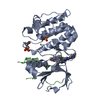
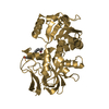
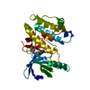
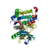
| Title | RMP-pepstatin A complex | |||||||||
 Components Components |
| |||||||||
 Keywords Keywords | HYDROLASE/HYDROLASE INHIBITOR / ASPARTIC PROTEINASE / PEPSTATIN A / ASPARTYL PROTEASE-PEPTIDE complex / HYDROLASE-HYDROLASE INHIBITOR complex | |||||||||
| Function / homology |  Function and homology information Function and homology information | |||||||||
| Biological species |  Rhizomucor miehei (fungus) Rhizomucor miehei (fungus) Streptomyces (bacteria) Streptomyces (bacteria) | |||||||||
| Method |  X-RAY DIFFRACTION / SYNCHROTRON / DIRECT ELECTRON DENSITY DIFFERENCE MAP / Resolution: 2.7 Å X-RAY DIFFRACTION / SYNCHROTRON / DIRECT ELECTRON DENSITY DIFFERENCE MAP / Resolution: 2.7 Å | |||||||||
 Authors Authors | Yang, J. / Quail, J.W. | |||||||||
 Citation Citation | Journal: Acta Crystallogr.,Sect.D / Year: 1999 Title: Structure of the Rhizomucor miehei aspartic proteinase complexed with the inhibitor pepstatin A at 2.7 A resolution. Authors: Yang, J. / Quail, J.W. | |||||||||
| History |
| |||||||||
| Remark 650 | HELIX DETERMINATION METHOD: AUTHOR-DETERMINED | |||||||||
| Remark 700 | SHEET DETERMINATION METHOD: TAKEN FROM RELEASED PDB ENTRY 2ASI |
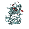
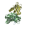
- Structure visualization
| Structure viewer | Molecule: MolmilJmol/JSmol |
|---|
- Downloads & links
-Download
| PDBx/mmCIF format | 2rmp.cif.gz | 83.2 KB | Display | PDBx/mmCIF format |
|---|---|---|---|---|
| PDB format | pdb2rmp.ent.gz | 61.8 KB | Display | PDB format |
| PDBx/mmJSON format | 2rmp.json.gz | Tree view | PDBx/mmJSON format | |
| Others |  Other downloads Other downloads |
-Validation report
| Arichive directory | https://data.pdbj.org/pub/pdb/validation_reports/rm/2rmpftp://data.pdbj.org/pub/pdb/validation_reports/rm/2rmp | HTTPS FTP |
|---|
-Related structure data
| Related structure data | 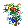 2asiS S: Starting model for refinement |
|---|---|
| Similar structure data |


-Links
 PDBj
PDBj
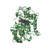
- Assembly
| Deposited unit | 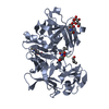
| ||||||||
|---|---|---|---|---|---|---|---|---|---|
| 1 |
| ||||||||
| Unit cell |
|
-Components
| #1: Protein | Mass: 38735.742 Da / Num. of mol.: 1 / Source method: isolated from a natural source / Source: (natural) Rhizomucor miehei (fungus) / References: UniProt: P00799, mucorpepsin |
|---|---|
| #2: Protein/peptide | 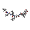  Type: Oligopeptide / Class: Enzyme inhibitor / Mass: 685.891 Da / Num. of mol.: 1 / Source method: isolated from a natural source Type: Oligopeptide / Class: Enzyme inhibitor / Mass: 685.891 Da / Num. of mol.: 1 / Source method: isolated from a natural sourceDetails: STREPTOMYCES (PEPSTATIN A WAS ISOLATED FROM STREPTOMYCES) Source: (natural) Streptomyces (bacteria) / Genus: Streptomyces / References: Pepstatin |
| #3: Polysaccharide | beta-D-mannopyranose-(1-4)-2-acetamido-2-deoxy-beta-D-glucopyranose Source method: isolated from a genetically manipulated source |
| #4: Sugar | ChemComp-NAG /   Type: D-saccharide, beta linking / Mass: 221.208 Da / Num. of mol.: 1 Type: D-saccharide, beta linking / Mass: 221.208 Da / Num. of mol.: 1Source method: isolated from a genetically manipulated source Formula: C8H15NO6 |
| Has protein modification | Y |
-Experimental details
-Experiment
| Experiment | Method: X-RAY DIFFRACTION / Number of used crystals: 1 |
|---|
- Sample preparation
| Crystal | Density Matthews: 2.31 Å3/Da / Density % sol: 49 % | ||||||||||||||||||||||||||||||
|---|---|---|---|---|---|---|---|---|---|---|---|---|---|---|---|---|---|---|---|---|---|---|---|---|---|---|---|---|---|---|---|
| Crystal grow | pH: 3.6 / Details: pH 3.6 | ||||||||||||||||||||||||||||||
| Crystal grow | *PLUS Method: vapor diffusion, hanging drop | ||||||||||||||||||||||||||||||
| Components of the solutions | *PLUS
|
-Data collection
| Diffraction | Mean temperature: 287 K |
|---|---|
| Diffraction source | Source: SYNCHROTRON / Site: NSLS  / Beamline: X12C / Wavelength: 1.15 / Beamline: X12C / Wavelength: 1.15 |
| Detector | Type: ENRAF-NONIUS FAST / Detector: DIFFRACTOMETER / Date: Nov 1, 1996 / Details: MIRROR |
| Radiation | Monochromator: SINGLE FLAT CRYSTALS / Monochromatic (M) / Laue (L): M / Scattering type: x-ray |
| Radiation wavelength | Wavelength: 1.15 Å / Relative weight: 1 |
| Reflection | Resolution: 2.7→50 Å / Num. obs: 9360 / % possible obs: 84.9 % / Observed criterion σ(I): 0 / Redundancy: 4.6 % / Biso Wilson estimate: 45.4 Å2 / Rsym value: 0.092 / Net I/σ(I): 15 |
| Reflection shell | Resolution: 2.7→2.8 Å / Redundancy: 2 % / Mean I/σ(I) obs: 2 / Rsym value: 0.159 / % possible all: 48 |
| Reflection | *PLUS Highest resolution: 2.7 Å / Num. measured all: 43156 / Rmerge(I) obs: 0.092 |
| Reflection shell | *PLUS Highest resolution: 2.7 Å / Lowest resolution: 2.8 Å / % possible obs: 47.9 % / Rmerge(I) obs: 0.159 |
- Processing
| Software |
| ||||||||||||||||||||||||||||||||||||||||||||||||||||||||||||
|---|---|---|---|---|---|---|---|---|---|---|---|---|---|---|---|---|---|---|---|---|---|---|---|---|---|---|---|---|---|---|---|---|---|---|---|---|---|---|---|---|---|---|---|---|---|---|---|---|---|---|---|---|---|---|---|---|---|---|---|---|---|
| Refinement | Method to determine structure: DIRECT ELECTRON DENSITY DIFFERENCE MAP Starting model: PDB ENTRY 2ASI Resolution: 2.7→8 Å / Rfactor Rfree error: 0.01 / Data cutoff high absF: 1000000 / Data cutoff low absF: 0.001 / Isotropic thermal model: RESTRAINED / Cross valid method: THROUGHOUT / σ(F): 0
| ||||||||||||||||||||||||||||||||||||||||||||||||||||||||||||
| Displacement parameters | Biso mean: 25.1 Å2 | ||||||||||||||||||||||||||||||||||||||||||||||||||||||||||||
| Refine analyze |
| ||||||||||||||||||||||||||||||||||||||||||||||||||||||||||||
| Refinement step | Cycle: LAST / Resolution: 2.7→8 Å
| ||||||||||||||||||||||||||||||||||||||||||||||||||||||||||||
| Refine LS restraints |
| ||||||||||||||||||||||||||||||||||||||||||||||||||||||||||||
| LS refinement shell | Resolution: 2.7→2.82 Å / Rfactor Rfree error: 0.06 / Total num. of bins used: 8
| ||||||||||||||||||||||||||||||||||||||||||||||||||||||||||||
| Xplor file |
| ||||||||||||||||||||||||||||||||||||||||||||||||||||||||||||
| Software | *PLUS Name: X-PLOR / Version: 3.8 / Classification: refinement | ||||||||||||||||||||||||||||||||||||||||||||||||||||||||||||
| Refinement | *PLUS Rfactor Rfree: 0.28 | ||||||||||||||||||||||||||||||||||||||||||||||||||||||||||||
| Solvent computation | *PLUS | ||||||||||||||||||||||||||||||||||||||||||||||||||||||||||||
| Displacement parameters | *PLUS | ||||||||||||||||||||||||||||||||||||||||||||||||||||||||||||
| Refine LS restraints | *PLUS
|
