- PDB-2rld: CRYSTAL STRUCTURE OF A PROTEIN WITH UNKNOWN FUNCTION FROM S23 RIB... -
+
Open data
ID or keywords:
Loading...
-
Basic information
Entry
Database: PDB / ID: 2rld
Title
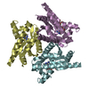
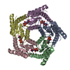
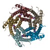
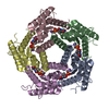
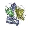
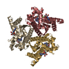
CRYSTAL STRUCTURE OF A PROTEIN WITH UNKNOWN FUNCTION FROM S23 RIBOSOMAL PROTEIN FAMILY (BT_0352) FROM BACTEROIDES THETAIOTAOMICRON VPI-5482 AT 1.70 A RESOLUTION
Components
Uncharacterized protein
Keywords
UNKNOWN FUNCTION / STRUCTURAL GENOMICS / JOINT CENTER FOR STRUCTURAL GENOMICS / JCSG / PROTEIN STRUCTURE INITIATIVE / PSI-2
Function / homology
23S rRNA-intervening sequence protein / 23S rRNA-intervening sequence protein / 23S rRNA-intervening sequence / 23S rRNA-intervening sequence superfamily / de novo design (two linked rop proteins) / Up-down Bundle / Mainly Alpha / metal ion binding / Four helix bundle protein
Function and homology information
Biological species
Bacteroides thetaiotaomicron VPI-5482 (bacteria)
Method
X-RAY DIFFRACTION / SYNCHROTRON / MAD / Resolution: 1.7 Å
SEQUENCE THE CONSTRUCT WAS EXPRESSED WITH A PURIFICATION TAG MGSDKIHHHHHHENLYFQG. THE TAG WAS ... SEQUENCE THE CONSTRUCT WAS EXPRESSED WITH A PURIFICATION TAG MGSDKIHHHHHHENLYFQG. THE TAG WAS REMOVED WITH TEV PROTEASE LEAVING ONLY A GLYCINE (0) FOLLOWED BY THE TARGET SEQUENCE.
A: Uncharacterized protein B: Uncharacterized protein C: Uncharacterized protein D: Uncharacterized protein E: Uncharacterized protein hetero molecules
Mass: 18.015 Da / Num. of mol.: 518 / Source method: isolated from a natural source / Formula: H2O
Has protein modification
Y
Sequence details
REMARK 999 SEQUENCE: THE CONSTRUCT WAS EXPRESSED WITH A PURIFICATION REMARK 999 TAG ...REMARK 999 SEQUENCE: THE CONSTRUCT WAS EXPRESSED WITH A PURIFICATION REMARK 999 TAG MGSDKIHHHHHHENLYFQG. THE TAG WAS REMOVED WITH TEV PROTEASE REMARK 999 LEAVING ONLY A GLYCINE (0) FOLLOWED BY THE TARGET SEQUENCE.
-
Experimental details
-
Experiment
Experiment
Method: X-RAY DIFFRACTION / Number of used crystals: 1
-
Sample preparation
Crystal
Density Matthews: 2.13 Å3/Da / Density % sol: 42.25 %
Crystal grow
Temperature: 277 K / Method: vapor diffusion, sitting drop / pH: 5.1 Details: NANODROP, 0.2M CaCl2, 20.0% PEG 3350, no Buffer pH 5.1, VAPOR DIFFUSION, SITTING DROP, temperature 277K
Resolution: 1.7→29.643 Å / Num. obs: 63158 / % possible obs: 96.2 % / Redundancy: 2 % / Biso Wilson estimate: 20.65 Å2 / Rmerge(I) obs: 0.053 / Rsym value: 0.053 / Net I/σ(I): 9.1
Reflection shell
Diffraction-ID: 1
Resolution (Å)
Redundancy (%)
Rmerge(I) obs
Mean I/σ(I) obs
Num. measured all
Num. unique all
Rsym value
% possible all
1.7-1.74
1.8
0.441
1.7
7791
4288
0.441
88.2
1.74-1.79
1.9
0.356
2.1
8308
4342
0.356
92.3
1.79-1.84
2
0.271
2.8
8702
4366
0.271
94.6
1.84-1.9
2
0.201
3.8
8731
4263
0.201
96.2
1.9-1.96
2.1
0.149
5
8754
4183
0.149
97
1.96-2.03
2.1
0.121
6.1
8607
4085
0.121
97.1
2.03-2.11
2.1
0.093
7.8
8236
3919
0.093
97.3
2.11-2.19
2.1
0.076
9.3
7976
3796
0.076
97.6
2.19-2.29
2.1
0.066
10.2
7624
3651
0.066
97.7
2.29-2.4
2.1
0.059
11.7
7240
3459
0.059
97.9
2.4-2.53
2.1
0.055
11.9
6930
3326
0.055
97.9
2.53-2.69
2.1
0.053
12.4
6545
3159
0.053
98
2.69-2.87
2.1
0.048
12.9
6057
2939
0.048
97.9
2.87-3.1
2.1
0.042
14.5
5724
2750
0.042
97.2
3.1-3.4
2
0.04
15.2
5162
2523
0.04
97.3
3.4-3.8
2
0.036
15.6
4545
2286
0.036
97.6
3.8-4.39
1.9
0.035
16.9
3714
1974
0.035
95.3
4.39-5.38
2.1
0.036
15.5
3616
1743
0.036
99
5.38-7.6
2.1
0.041
15
2889
1366
0.041
99.8
7.6-29.643
2.1
0.033
17.4
1551
740
0.033
96.4
-
Phasing
Phasing
Method: MAD
-
Processing
Software
Name
Version
Classification
NB
REFMAC
5.2.0019
refinement
PHENIX
refinement
SHELX
phasing
MolProbity
3beta29
modelbuilding
SCALA
datascaling
PDB_EXTRACT
3
dataextraction
ADSC
Quantum
datacollection
MOSFLM
datareduction
SHELXD
phasing
SOLVE
phasing
Refinement
Method to determine structure: MAD / Resolution: 1.7→29.643 Å / Cor.coef. Fo:Fc: 0.965 / Cor.coef. Fo:Fc free: 0.949 / SU B: 4.051 / SU ML: 0.067 / TLS residual ADP flag: LIKELY RESIDUAL / Cross valid method: THROUGHOUT / σ(F): 0 / ESU R: 0.104 / ESU R Free: 0.104 Stereochemistry target values: MAXIMUM LIKELIHOOD WITH PHASES Details: 1. HYDROGENS HAVE BEEN ADDED IN THE RIDING POSITIONS. 2. ATOM RECORDS CONTAIN RESIDUAL B FACTORS ONLY. 3. A MET-INHIBITION PROTOCOL WAS USED FOR SELENOMETHIONINE INCORPORATION DURING PROTEIN ...Details: 1. HYDROGENS HAVE BEEN ADDED IN THE RIDING POSITIONS. 2. ATOM RECORDS CONTAIN RESIDUAL B FACTORS ONLY. 3. A MET-INHIBITION PROTOCOL WAS USED FOR SELENOMETHIONINE INCORPORATION DURING PROTEIN EXPRESSION. THE OCCUPANCY OF THE SE ATOMS IN THE MSE RESIDUES WAS REDUCED TO 0.75 FOR THE REDUCED SCATTERING POWER DUE TO PARTIAL S-MET INCORPORATION. 4. EDO, CL AND CA MOLECULES FROM THE CRYSTALLIZATION/CRYO SOLUTION ARE MODELED.
Rfactor
Num. reflection
% reflection
Selection details
Rfree
0.201
3216
5.1 %
RANDOM
Rwork
0.163
-
-
-
obs
0.165
63130
95.92 %
-
Solvent computation
Ion probe radii: 0.8 Å / Shrinkage radii: 0.8 Å / VDW probe radii: 1.2 Å / Solvent model: BABINET MODEL WITH MASK
In the structure databanks used in Yorodumi, some data are registered as the other names, "COVID-19 virus" and "2019-nCoV". Here are the details of the virus and the list of structure data.
Jan 31, 2019. EMDB accession codes are about to change! (news from PDBe EMDB page)
EMDB accession codes are about to change! (news from PDBe EMDB page)
The allocation of 4 digits for EMDB accession codes will soon come to an end. Whilst these codes will remain in use, new EMDB accession codes will include an additional digit and will expand incrementally as the available range of codes is exhausted. The current 4-digit format prefixed with “EMD-” (i.e. EMD-XXXX) will advance to a 5-digit format (i.e. EMD-XXXXX), and so on. It is currently estimated that the 4-digit codes will be depleted around Spring 2019, at which point the 5-digit format will come into force.
The EM Navigator/Yorodumi systems omit the EMD- prefix.
Related info.:Q: What is EMD? / ID/Accession-code notation in Yorodumi/EM Navigator
Yorodumi is a browser for structure data from EMDB, PDB, SASBDB, etc.
This page is also the successor to EM Navigator detail page, and also detail information page/front-end page for Omokage search.
The word "yorodu" (or yorozu) is an old Japanese word meaning "ten thousand". "mi" (miru) is to see.
Related info.:EMDB / PDB / SASBDB / Comparison of 3 databanks / Yorodumi Search / Aug 31, 2016. New EM Navigator & Yorodumi / Yorodumi Papers / Jmol/JSmol / Function and homology information / Changes in new EM Navigator and Yorodumi
 Movie
Movie Controller

 Yorodumi
 Open data
 Basic information
 Components
 Keywords
 Function and homology information
 Bacteroides thetaiotaomicron VPI-5482 (bacteria)
 X-RAY DIFFRACTION /
  Authors
 Citation
 Structure visualization
 Downloads & links
 Other downloads
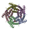

 PDBj
PDBj Assembly

 Mass: 40.078 Da / Num. of mol.: 3 / Source method: obtained synthetically / Formula: Ca
 Mass: 35.453 Da / Num. of mol.: 2 / Source method: obtained synthetically / Formula: Cl
 Mass: 62.068 Da / Num. of mol.: 16 / Source method: obtained synthetically / Formula: C2H6O2
 Mass: 18.015 Da / Num. of mol.: 518 / Source method: isolated from a natural source / Formula: H2O
 Sample preparation
 / Beamline: 8.2.2 / Wavelength: 0.9798, 0.9537, 0.9796
 Processing