 Movie
Movie Controller


+ Open data

- Basic information
| Entry | Database: PDB / ID: 2rk3 | ||||||
|---|---|---|---|---|---|---|---|
| Title | Structure of A104T DJ-1 | ||||||
 Components Components | Protein DJ-1 | ||||||
 Keywords Keywords | CHAPERONE / PARKINSON'S DISEASE / THIJ / PFPI / Cytoplasm / Disease mutation / Nucleus / Oncogene / Oxidation / Parkinson disease / Phosphorylation / Polymorphism / Ubl conjugation | ||||||
| Function / homology |  Function and homology information Function and homology informationpositive regulation of acute inflammatory response to antigenic stimulus / tyrosine 3-monooxygenase activator activity / cellular response to glyoxal / L-dopa decarboxylase activator activity / detoxification of hydrogen peroxide / : / detection of oxidative stress / : / guanine deglycation, glyoxal removal / cellular detoxification of methylglyoxal ...positive regulation of acute inflammatory response to antigenic stimulus / tyrosine 3-monooxygenase activator activity / cellular response to glyoxal / L-dopa decarboxylase activator activity / detoxification of hydrogen peroxide / : / detection of oxidative stress / : / guanine deglycation, glyoxal removal / cellular detoxification of methylglyoxal / regulation of supramolecular fiber organization / negative regulation of death-inducing signaling complex assembly / negative regulation of TRAIL-activated apoptotic signaling pathway / : / glyoxalase (glycolic acid-forming) activity / negative regulation of protein K48-linked deubiquitination / negative regulation of nitrosative stress-induced intrinsic apoptotic signaling pathway / glycolate biosynthetic process / glyoxal metabolic process / guanine deglycation / detoxification of mercury ion / ubiquitin-protein transferase inhibitor activity / protein deglycase / hydrogen peroxide metabolic process / mercury ion binding / methylglyoxal metabolic process / protein deglycase activity / positive regulation of autophagy of mitochondrion / superoxide dismutase copper chaperone activity / oxidoreductase activity, acting on peroxide as acceptor / positive regulation of dopamine biosynthetic process / positive regulation of mitochondrial electron transport, NADH to ubiquinone / lactate biosynthetic process / negative regulation of hydrogen peroxide-induced neuron intrinsic apoptotic signaling pathway / protein repair / peptidase inhibitor activity / peroxiredoxin activity / cellular detoxification of aldehyde / small protein activating enzyme binding / Hydrolases; Acting on ester bonds; Thioester hydrolases / regulation of oxidative stress-induced neuron intrinsic apoptotic signaling pathway / detoxification of copper ion / negative regulation of protein sumoylation / negative regulation of protein export from nucleus / cupric ion binding / negative regulation of oxidative stress-induced neuron intrinsic apoptotic signaling pathway / regulation of androgen receptor signaling pathway / insulin secretion / Hydrolases; Acting on carbon-nitrogen bonds, other than peptide bonds; In linear amides / oxygen sensor activity / androgen receptor signaling pathway / nuclear androgen receptor binding / negative regulation of intrinsic apoptotic signaling pathway in response to hydrogen peroxide / ubiquitin-like protein conjugating enzyme binding / single fertilization / ubiquitin-specific protease binding / cytokine binding / cuprous ion binding / signaling receptor activator activity / regulation of synaptic vesicle endocytosis / negative regulation of endoplasmic reticulum stress-induced intrinsic apoptotic signaling pathway / negative regulation of oxidative stress-induced intrinsic apoptotic signaling pathway / negative regulation of reactive oxygen species biosynthetic process / removal of superoxide radicals / SUMOylation of transcription cofactors / negative regulation of proteasomal ubiquitin-dependent protein catabolic process / negative regulation of protein ubiquitination / regulation of neuron apoptotic process / negative regulation of extrinsic apoptotic signaling pathway / regulation of mitochondrial membrane potential / positive regulation of interleukin-8 production / adherens junction / mitochondrion organization / positive regulation of protein-containing complex assembly / Late endosomal microautophagy / positive regulation of protein localization to nucleus / mitochondrial intermembrane space / PML body / autophagy / positive regulation of reactive oxygen species metabolic process / cellular response to hydrogen peroxide / enzyme activator activity / kinase binding / Chaperone Mediated Autophagy / Aggrephagy / peptidase activity / glucose homeostasis / synaptic vesicle / regulation of inflammatory response / cellular response to oxidative stress / cell body / response to oxidative stress / scaffold protein binding / DNA-binding transcription factor binding / negative regulation of neuron apoptotic process / Ras protein signal transduction / transcription coactivator activity / positive regulation of phosphatidylinositol 3-kinase/protein kinase B signal transduction / protein stabilization / cadherin binding Similarity search - Function | ||||||
| Biological species |  Homo sapiens (human) Homo sapiens (human) | ||||||
| Method |  X-RAY DIFFRACTION / SYNCHROTRON / MOLECULAR REPLACEMENT / Resolution: 1.05 Å X-RAY DIFFRACTION / SYNCHROTRON / MOLECULAR REPLACEMENT / Resolution: 1.05 Å | ||||||
 Authors Authors | Lakshminarasimhan, M. / Maldonado, M.T. / Zhou, W. / Fink, A.L. / Wilson, M.A. | ||||||
 Citation Citation | Journal: Biochemistry / Year: 2008 Title: Structural Impact of Three Parkinsonism-Associated Missense Mutations on Human DJ-1. Authors: Lakshminarasimhan, M. / Maldonado, M.T. / Zhou, W. / Fink, A.L. / Wilson, M.A. | ||||||
| History |
|
- Structure visualization
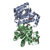
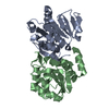
| Structure viewer | Molecule: MolmilJmol/JSmol |
|---|
- Downloads & links
-Download
| PDBx/mmCIF format | 2rk3.cif.gz | 94.9 KB | Display | PDBx/mmCIF format |
|---|---|---|---|---|
| PDB format | pdb2rk3.ent.gz | 71.7 KB | Display | PDB format |
| PDBx/mmJSON format | 2rk3.json.gz | Tree view | PDBx/mmJSON format | |
| Others |  Other downloads Other downloads |
-Validation report
| Arichive directory | https://data.pdbj.org/pub/pdb/validation_reports/rk/2rk3ftp://data.pdbj.org/pub/pdb/validation_reports/rk/2rk3 | HTTPS FTP |
|---|
-Related structure data
| Related structure data |  2rk4C  2rk6C  3b36C  3b38C  3b3aC  1p5fS C: citing same article ( S: Starting model for refinement |
|---|---|
| Similar structure data |


-Links
 PDBj
PDBj


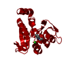
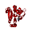
- Assembly
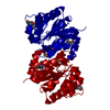
| Deposited unit | 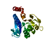
| ||||||||
|---|---|---|---|---|---|---|---|---|---|
| 1 | 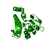
| ||||||||
| Unit cell |
| ||||||||
| Components on special symmetry positions |
|
-Components
| #1: Protein | Mass: 21018.232 Da / Num. of mol.: 1 / Mutation: A104T Source method: isolated from a genetically manipulated source Source: (gene. exp.) Homo sapiens (human) / Gene: PARK7 / Plasmid: pET15b / Species (production host): Escherichia coli / Production host:  |
|---|---|
| #2: Water | ChemComp-HOH /  Mass: 18.015 Da / Num. of mol.: 229 / Source method: isolated from a natural source / Formula: H2O Mass: 18.015 Da / Num. of mol.: 229 / Source method: isolated from a natural source / Formula: H2O |
-Experimental details
-Experiment
| Experiment | Method: X-RAY DIFFRACTION / Number of used crystals: 1 |
|---|
- Sample preparation
| Crystal | Density Matthews: 3.03 Å3/Da / Density % sol: 59.46 % |
|---|---|
| Crystal grow | Temperature: 298 K / Method: vapor diffusion, hanging drop / pH: 7.5 Details: 30% PEG 3000, 100 mM HEPES, 200 mM NaCl, pH 7.5, VAPOR DIFFUSION, HANGING DROP, temperature 298K |
-Data collection
| Diffraction | Mean temperature: 110 K |
|---|---|
| Diffraction source | Source: SYNCHROTRON / Site: SSRL  / Beamline: BL11-1 / Wavelength: 0.98 Å / Beamline: BL11-1 / Wavelength: 0.98 Å |
| Detector | Type: ADSC QUANTUM 315 / Detector: CCD / Date: Apr 25, 2005 |
| Radiation | Monochromator: Single Crystal Bent Si(111) / Protocol: SINGLE WAVELENGTH / Monochromatic (M) / Laue (L): M / Scattering type: x-ray |
| Radiation wavelength | Wavelength: 0.98 Å / Relative weight: 1 |
| Reflection | Resolution: 1.05→50 Å / Num. all: 110339 / Num. obs: 110339 / % possible obs: 97.8 % / Redundancy: 5.7 % / Rmerge(I) obs: 0.043 / Net I/σ(I): 33.9 |
| Reflection shell | Resolution: 1.05→1.09 Å / Redundancy: 3.8 % / Rmerge(I) obs: 0.437 / Mean I/σ(I) obs: 2.6 / Num. unique all: 10567 / % possible all: 94.5 |
- Processing
| Software |
| |||||||||||||||||||||||||||||||||
|---|---|---|---|---|---|---|---|---|---|---|---|---|---|---|---|---|---|---|---|---|---|---|---|---|---|---|---|---|---|---|---|---|---|---|
| Refinement | Method to determine structure: MOLECULAR REPLACEMENT Starting model: 1P5F Resolution: 1.05→50 Å / Num. parameters: 15298 / Num. restraintsaints: 19492 / Cross valid method: FREE R / σ(F): 0 / Stereochemistry target values: Engh & Huber
| |||||||||||||||||||||||||||||||||
| Solvent computation | Solvent model: BABINET | |||||||||||||||||||||||||||||||||
| Refine analyze | Num. disordered residues: 12 / Occupancy sum hydrogen: 1409 / Occupancy sum non hydrogen: 1604 | |||||||||||||||||||||||||||||||||
| Refinement step | Cycle: LAST / Resolution: 1.05→50 Å
| |||||||||||||||||||||||||||||||||
| Refine LS restraints |
|
