- PDB-2rjo: Crystal structure of Twin-arginine translocation pathway signal p... -
+
Open data
ID or keywords:
Loading...
-
Basic information
Entry
Database: PDB / ID: 2rjo
Title
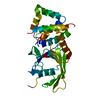
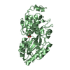
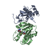
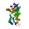
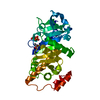
Crystal structure of Twin-arginine translocation pathway signal protein from Burkholderia phytofirmans
Components
Twin-arginine translocation pathway signal protein
Keywords
SIGNALING PROTEIN / PSI-2 / NYSGXRC / twin arginine translocation pathway signal protein / Structural Genomics / Protein Structure Initiative / New York SGX Research Center for Structural Genomics
Monochromator: Si-III / Protocol: SINGLE WAVELENGTH / Monochromatic (M) / Laue (L): M / Scattering type: x-ray
Radiation wavelength
Wavelength: 0.979 Å / Relative weight: 1
Reflection
Resolution: 2.04→50 Å / Num. all: 20727 / Num. obs: 20727 / % possible obs: 85.1 % / Observed criterion σ(F): 0 / Redundancy: 10.2 % / Biso Wilson estimate: 12.7 Å2 / Rmerge(I) obs: 0.07 / Net I/σ(I): 8.6
Reflection shell
Resolution: 2.04→2.11 Å / Redundancy: 2.9 % / Rmerge(I) obs: 0.351 / Mean I/σ(I) obs: 1 / Num. unique all: 1039 / % possible all: 44.2
-
Processing
Software
Name
Version
Classification
CNS
1.1
refinement
CBASS
datacollection
HKL-2000
datacollection
HKL-2000
datareduction
HKL-2000
datascaling
SHELXD
phasing
SHELXE
modelbuilding
CCP4
phasing
Refinement
Method to determine structure: SAD / Resolution: 2.05→41.2 Å / Rfactor Rfree error: 0.012 / Data cutoff high absF: 57664.08 / Data cutoff low absF: 0 / Isotropic thermal model: RESTRAINED / Stereochemistry target values: Engh & Huber Details: Residues listed as missing in Remark 465 are due to lack of electron density. Residues with missing atoms listed in Remark 470 are due to lack of electron density for side chains and modeled as alanines.
Rfactor
Num. reflection
% reflection
Selection details
Rfree
0.281
560
2.9 %
RANDOM
Rwork
0.234
-
-
-
obs
0.234
19564
82.3 %
-
all
-
20727
-
-
Solvent computation
Bsol: 38.5128 Å2 / ksol: 0.372992 e/Å3
Displacement parameters
Biso mean: 25.2 Å2
Baniso -1
Baniso -2
Baniso -3
1-
10.92 Å2
0 Å2
0 Å2
2-
-
-6.58 Å2
0 Å2
3-
-
-
-4.34 Å2
Refine analyze
Luzzati d res low obs: 5 Å
Refinement step
Cycle: LAST / Resolution: 2.05→41.2 Å
Protein
Nucleic acid
Ligand
Solvent
Total
Num. atoms
2438
0
32
119
2589
Refine LS restraints
Refine-ID
Type
Dev ideal
X-RAY DIFFRACTION
c_bond_d
0.009
X-RAY DIFFRACTION
c_angle_deg
1.3
X-RAY DIFFRACTION
c_dihedral_angle_d
23.1
X-RAY DIFFRACTION
c_improper_angle_d
0.81
LS refinement shell
Resolution: 2.05→2.18 Å / Rfactor Rfree error: 0.041 / Total num. of bins used: 6
Rfactor
Num. reflection
% reflection
Rfree
0.299
52
2.7 %
Rwork
0.322
1848
-
obs
-
-
49.2 %
Xplor file
Refine-ID
Serial no
Topol file
X-RAY DIFFRACTION
1
protein.top
X-RAY DIFFRACTION
2
gal.top
+
About Yorodumi
-
News
-
Feb 9, 2022. New format data for meta-information of EMDB entries
New format data for meta-information of EMDB entries
Version 3 of the EMDB header file is now the official format.
The previous official version 1.9 will be removed from the archive.
In the structure databanks used in Yorodumi, some data are registered as the other names, "COVID-19 virus" and "2019-nCoV". Here are the details of the virus and the list of structure data.
Jan 31, 2019. EMDB accession codes are about to change! (news from PDBe EMDB page)
EMDB accession codes are about to change! (news from PDBe EMDB page)
The allocation of 4 digits for EMDB accession codes will soon come to an end. Whilst these codes will remain in use, new EMDB accession codes will include an additional digit and will expand incrementally as the available range of codes is exhausted. The current 4-digit format prefixed with “EMD-” (i.e. EMD-XXXX) will advance to a 5-digit format (i.e. EMD-XXXXX), and so on. It is currently estimated that the 4-digit codes will be depleted around Spring 2019, at which point the 5-digit format will come into force.
The EM Navigator/Yorodumi systems omit the EMD- prefix.
Related info.:Q: What is EMD? / ID/Accession-code notation in Yorodumi/EM Navigator
Yorodumi is a browser for structure data from EMDB, PDB, SASBDB, etc.
This page is also the successor to EM Navigator detail page, and also detail information page/front-end page for Omokage search.
The word "yorodu" (or yorozu) is an old Japanese word meaning "ten thousand". "mi" (miru) is to see.
Related info.:EMDB / PDB / SASBDB / Comparison of 3 databanks / Yorodumi Search / Aug 31, 2016. New EM Navigator & Yorodumi / Yorodumi Papers / Jmol/JSmol / Function and homology information / Changes in new EM Navigator and Yorodumi
 Movie
Movie Controller

 Yorodumi
 Open data
 Basic information
 Components
 Keywords
 Function and homology information
 Burkholderia phytofirmans (bacteria)
 X-RAY DIFFRACTION /
  Authors
 Citation
 Structure visualization
 Downloads & links
 Other downloads
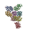


 PDBj
PDBj


 Assembly
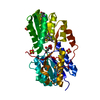

 Type: D-saccharide, beta linking / Mass: 180.156 Da / Num. of mol.: 1
 Mass: 96.063 Da / Num. of mol.: 4 / Source method: obtained synthetically / Formula: SO4
 Mass: 18.015 Da / Num. of mol.: 119 / Source method: isolated from a natural source / Formula: H2O
 Sample preparation
 / Beamline: X29A / Wavelength: 0.979 Å
 Processing