Monochromator: double crystal / Protocol: SINGLE WAVELENGTH / Monochromatic (M) / Laue (L): M / Scattering type: x-ray
Radiation wavelength
Wavelength: 0.97918 Å / Relative weight: 1
Reflection
Resolution: 2.6→50 Å / Num. all: 30250 / Num. obs: 28834 / % possible obs: 95.3 % / Observed criterion σ(I): -3 / Redundancy: 5.3 % / Biso Wilson estimate: 59.191 Å2 / Rmerge(I) obs: 0.095 / Net I/σ(I): 35.58
Reflection shell
Resolution: 2.6→2.62 Å / Redundancy: 4.4 % / Rmerge(I) obs: 0.256 / Mean I/σ(I) obs: 6.19 / Num. unique all: 319 / % possible all: 78.6
-
Processing
Software
Name
Version
Classification
REFMAC
5.2.0019
refinement
SBC-Collect
datacollection
HKL-3000
datareduction
HKL-3000
datascaling
HKL-3000
phasing
Refinement
Method to determine structure: SAD / Resolution: 2.6→41.03 Å / Cor.coef. Fo:Fc: 0.945 / Cor.coef. Fo:Fc free: 0.907 / SU B: 14.271 / SU ML: 0.262 / Cross valid method: THROUGHOUT / σ(F): 0 / ESU R: 0.67 / ESU R Free: 0.328 / Stereochemistry target values: MAXIMUM LIKELIHOOD Details: The Bijvoet differences were used for phasing. HYDROGENS HAVE BEEN ADDED IN THE RIDING POSITIONS
Rfactor
Num. reflection
% reflection
Selection details
Rfree
0.26533
736
5 %
RANDOM
Rwork
0.20846
-
-
-
all
0.21124
13868
-
-
obs
0.21124
13868
96.48 %
-
Solvent computation
Ion probe radii: 0.8 Å / Shrinkage radii: 0.8 Å / VDW probe radii: 1.2 Å / Solvent model: MASK
Displacement parameters
Biso mean: 65.333 Å2
Baniso -1
Baniso -2
Baniso -3
1-
-3.78 Å2
-1.89 Å2
0 Å2
2-
-
-3.78 Å2
0 Å2
3-
-
-
5.67 Å2
Refinement step
Cycle: LAST / Resolution: 2.6→41.03 Å
Protein
Nucleic acid
Ligand
Solvent
Total
Num. atoms
2908
0
0
48
2956
Refine LS restraints
Refine-ID
Type
Dev ideal
Dev ideal target
Number
X-RAY DIFFRACTION
r_bond_refined_d
0.012
0.021
2957
X-RAY DIFFRACTION
r_bond_other_d
X-RAY DIFFRACTION
r_angle_refined_deg
1.353
1.957
3996
X-RAY DIFFRACTION
r_angle_other_deg
X-RAY DIFFRACTION
r_dihedral_angle_1_deg
5.912
5
379
X-RAY DIFFRACTION
r_dihedral_angle_2_deg
36.047
24.18
122
X-RAY DIFFRACTION
r_dihedral_angle_3_deg
15.71
15
508
X-RAY DIFFRACTION
r_dihedral_angle_4_deg
17.252
15
17
X-RAY DIFFRACTION
r_chiral_restr
0.094
0.2
475
X-RAY DIFFRACTION
r_gen_planes_refined
0.004
0.02
2179
X-RAY DIFFRACTION
r_gen_planes_other
X-RAY DIFFRACTION
r_nbd_refined
0.226
0.2
1301
X-RAY DIFFRACTION
r_nbd_other
X-RAY DIFFRACTION
r_nbtor_refined
0.302
0.2
2024
X-RAY DIFFRACTION
r_nbtor_other
X-RAY DIFFRACTION
r_xyhbond_nbd_refined
0.146
0.2
91
X-RAY DIFFRACTION
r_xyhbond_nbd_other
X-RAY DIFFRACTION
r_metal_ion_refined
X-RAY DIFFRACTION
r_metal_ion_other
X-RAY DIFFRACTION
r_symmetry_vdw_refined
0.266
0.2
26
X-RAY DIFFRACTION
r_symmetry_vdw_other
X-RAY DIFFRACTION
r_symmetry_hbond_refined
0.342
0.2
7
X-RAY DIFFRACTION
r_symmetry_hbond_other
X-RAY DIFFRACTION
r_symmetry_metal_ion_refined
X-RAY DIFFRACTION
r_symmetry_metal_ion_other
X-RAY DIFFRACTION
r_mcbond_it
0.931
1.5
1952
X-RAY DIFFRACTION
r_mcbond_other
X-RAY DIFFRACTION
r_mcangle_it
1.463
2
3048
X-RAY DIFFRACTION
r_scbond_it
2.364
3
1086
X-RAY DIFFRACTION
r_scangle_it
3.559
4.5
948
X-RAY DIFFRACTION
r_rigid_bond_restr
X-RAY DIFFRACTION
r_sphericity_free
X-RAY DIFFRACTION
r_sphericity_bonded
LS refinement shell
Resolution: 2.6→2.665 Å / Total num. of bins used: 20
Rfactor
Num. reflection
% reflection
Rfree
0.38
52
-
Rwork
0.265
849
-
obs
-
-
78.35 %
Refinement TLS params.
Method: refined / Refine-ID: X-RAY DIFFRACTION
ID
L11 (°2)
L12 (°2)
L13 (°2)
L22 (°2)
L23 (°2)
L33 (°2)
S11 (Å °)
S12 (Å °)
S13 (Å °)
S21 (Å °)
S22 (Å °)
S23 (Å °)
S31 (Å °)
S32 (Å °)
S33 (Å °)
T11 (Å2)
T12 (Å2)
T13 (Å2)
T22 (Å2)
T23 (Å2)
T33 (Å2)
Origin x (Å)
Origin y (Å)
Origin z (Å)
1
11.6039
4.4575
0.4504
2.7013
-0.0452
2.3248
0.0608
-0.4007
-0.8592
0.2597
-0.1776
-0.5801
0.6021
-0.3422
0.1168
-0.1786
-0.1335
-0.0346
-0.1595
0.0454
0.0908
29.8747
75.9784
44.1561
2
7.1038
5.1535
-2.8462
4.0188
-1.3178
3.1326
0.2528
0.1416
0.9511
0.0007
-0.2112
0.8357
-0.0408
-0.5684
-0.0416
-0.3004
-0.1066
0.023
0.103
-0.0357
0.2379
18.6244
85.98
41.1457
3
6.3755
3.6805
-0.0359
3.4212
-0.9607
0.6817
0.1941
-0.1471
-1.0858
0.2023
-0.2735
-0.7403
0.1596
-0.2733
0.0794
-0.1358
-0.0849
0.0105
-0.0349
0.0062
0.2363
35.3492
79.3657
43.0367
Refinement TLS group
ID
Refine-ID
Refine TLS-ID
Auth asym-ID
Label asym-ID
Auth seq-ID
Label seq-ID
1
X-RAY DIFFRACTION
1
A
A
4 - 14
7 - 17
2
X-RAY DIFFRACTION
1
A
A
51 - 61
54 - 64
3
X-RAY DIFFRACTION
1
A
A
65 - 75
68 - 78
4
X-RAY DIFFRACTION
1
A
A
103 - 118
106 - 121
5
X-RAY DIFFRACTION
1
A
A
132 - 181
135 - 184
6
X-RAY DIFFRACTION
2
B
B
16 - 50
19 - 53
7
X-RAY DIFFRACTION
2
B
B
81 - 102
84 - 105
8
X-RAY DIFFRACTION
3
B
B
119 - 131
122 - 134
9
X-RAY DIFFRACTION
3
B
B
182 - 203
185 - 206
+
About Yorodumi
-
News
-
Feb 9, 2022. New format data for meta-information of EMDB entries
New format data for meta-information of EMDB entries
Version 3 of the EMDB header file is now the official format.
The previous official version 1.9 will be removed from the archive.
In the structure databanks used in Yorodumi, some data are registered as the other names, "COVID-19 virus" and "2019-nCoV". Here are the details of the virus and the list of structure data.
Jan 31, 2019. EMDB accession codes are about to change! (news from PDBe EMDB page)
EMDB accession codes are about to change! (news from PDBe EMDB page)
The allocation of 4 digits for EMDB accession codes will soon come to an end. Whilst these codes will remain in use, new EMDB accession codes will include an additional digit and will expand incrementally as the available range of codes is exhausted. The current 4-digit format prefixed with “EMD-” (i.e. EMD-XXXX) will advance to a 5-digit format (i.e. EMD-XXXXX), and so on. It is currently estimated that the 4-digit codes will be depleted around Spring 2019, at which point the 5-digit format will come into force.
The EM Navigator/Yorodumi systems omit the EMD- prefix.
Related info.:Q: What is EMD? / ID/Accession-code notation in Yorodumi/EM Navigator
Yorodumi is a browser for structure data from EMDB, PDB, SASBDB, etc.
This page is also the successor to EM Navigator detail page, and also detail information page/front-end page for Omokage search.
The word "yorodu" (or yorozu) is an old Japanese word meaning "ten thousand". "mi" (miru) is to see.
Related info.:EMDB / PDB / SASBDB / Comparison of 3 databanks / Yorodumi Search / Aug 31, 2016. New EM Navigator & Yorodumi / Yorodumi Papers / Jmol/JSmol / Function and homology information / Changes in new EM Navigator and Yorodumi
 Movie
Movie Controller


 Open data
 Basic information
 Components
 Keywords
 Function and homology information
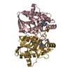
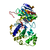
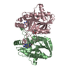
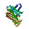
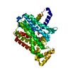
 Vibrio cholerae O1 biovar eltor str. N16961 (bacteria)
 X-RAY DIFFRACTION /
  Authors
 Citation
 Structure visualization
 Downloads & links
 Other downloads
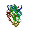


 PDBj
PDBj

 Assembly

 Mass: 60.052 Da / Num. of mol.: 2 / Source method: obtained synthetically / Formula: C2H4O2
 Mass: 18.015 Da / Num. of mol.: 40 / Source method: isolated from a natural source / Formula: H2O
 Sample preparation
 / Beamline: 19-ID / Wavelength: 0.97918 Å
 Processing