 Movie
Movie Controller

[English] 日本語
 Yorodumi
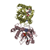
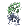
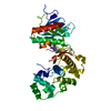
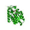
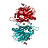
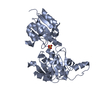
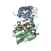
- PDB-2qtr: Crystal Structure of Nicotinate Mononucleotide Adenylyltransferase -
+ Open data

- Basic information
| Entry | Database: PDB / ID: 2qtr | |||||||||
|---|---|---|---|---|---|---|---|---|---|---|
| Title | Crystal Structure of Nicotinate Mononucleotide Adenylyltransferase | |||||||||
 Components Components | Probable nicotinate-nucleotide adenylyltransferase | |||||||||
 Keywords Keywords | TRANSFERASE / NAD / Nucleotidyltransferase / Pyridine nucleotide biosynthesis | |||||||||
| Function / homology |  Function and homology information Function and homology informationnicotinate-nucleotide adenylyltransferase / nicotinate-nucleotide adenylyltransferase activity / NAD+ biosynthetic process / ATP binding Similarity search - Function | |||||||||
| Biological species |  | |||||||||
| Method |  X-RAY DIFFRACTION / SYNCHROTRON / MOLECULAR REPLACEMENT / Resolution: 1.7 Å X-RAY DIFFRACTION / SYNCHROTRON / MOLECULAR REPLACEMENT / Resolution: 1.7 Å | |||||||||
 Authors Authors | Sershon, V.C. / Santarsiero, B.D. / Mesecar, A.D. | |||||||||
 Citation Citation | Journal: J.Mol.Biol. / Year: 2009 Title: Kinetic and X-ray structural evidence for negative cooperativity in substrate binding to nicotinate mononucleotide adenylyltransferase (NMAT) from Bacillus anthracis. Authors: Sershon, V.C. / Santarsiero, B.D. / Mesecar, A.D. | |||||||||
| History |
|
- Structure visualization
| Structure viewer | Molecule: MolmilJmol/JSmol |
|---|
- Downloads & links
-Download
| PDBx/mmCIF format | 2qtr.cif.gz | 140.7 KB | Display | PDBx/mmCIF format |
|---|---|---|---|---|
| PDB format | pdb2qtr.ent.gz | 111.1 KB | Display | PDB format |
| PDBx/mmJSON format | 2qtr.json.gz | Tree view | PDBx/mmJSON format | |
| Others |  Other downloads Other downloads |
-Validation report
| Summary document | 2qtr_validation.pdf.gz | 1.2 MB | Display | wwPDB validaton report |
|---|---|---|---|---|
| Full document | 2qtr_full_validation.pdf.gz | 1.2 MB | Display | |
| Data in XML | 2qtr_validation.xml.gz | 28.8 KB | Display | |
| Data in CIF | 2qtr_validation.cif.gz | 41.1 KB | Display | |
| Arichive directory | https://data.pdbj.org/pub/pdb/validation_reports/qt/2qtrftp://data.pdbj.org/pub/pdb/validation_reports/qt/2qtr | HTTPS FTP |
-Related structure data
| Related structure data | 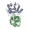 2qtmC 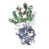 2qtnC 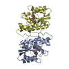 1kaqS C: citing same article ( S: Starting model for refinement |
|---|---|
| Similar structure data |


-Links
 PDBj
PDBj- Assembly
| Deposited unit | 
| ||||||||||||||||||
|---|---|---|---|---|---|---|---|---|---|---|---|---|---|---|---|---|---|---|---|
| 1 | 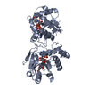
| ||||||||||||||||||
| 2 | 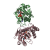
| ||||||||||||||||||
| Unit cell |
| ||||||||||||||||||
| Components on special symmetry positions |
| ||||||||||||||||||
| Details | BIOLOGICAL UNIT IS A DIMER OF CHAINS B AND C IN THE ASYMMETRIC UNIT, OR CHAIN A WITH A TWO-FOLD SYMMETRY MATE A' |
-Components
| #1: Protein | Mass: 21982.320 Da / Num. of mol.: 3 Source method: isolated from a genetically manipulated source Source: (gene. exp.) References: UniProt: A0A2B6C295, UniProt: C3L5T6*PLUS, nicotinate-nucleotide adenylyltransferase #2: Chemical | ChemComp-SO4 /   Mass: 96.063 Da / Num. of mol.: 4 / Source method: obtained synthetically / Formula: SO4 Mass: 96.063 Da / Num. of mol.: 4 / Source method: obtained synthetically / Formula: SO4#3: Chemical |   Mass: 665.418 Da / Num. of mol.: 3 / Source method: obtained synthetically / Formula: C21H27N6O15P2 Mass: 665.418 Da / Num. of mol.: 3 / Source method: obtained synthetically / Formula: C21H27N6O15P2#4: Water | ChemComp-HOH / |  Mass: 18.015 Da / Num. of mol.: 417 / Source method: isolated from a natural source / Formula: H2O Mass: 18.015 Da / Num. of mol.: 417 / Source method: isolated from a natural source / Formula: H2O |
|---|
-Experimental details
-Experiment
| Experiment | Method: X-RAY DIFFRACTION / Number of used crystals: 1 |
|---|
- Sample preparation
| Crystal | Density Matthews: 2.43 Å3/Da / Density % sol: 49.34 % |
|---|---|
| Crystal grow | Temperature: 295 K / Method: vapor diffusion, hanging drop / pH: 7.2 Details: sodium chloride, ATP, nicotinc acid, MgCl2, lithium sulfate, pH 7.2, VAPOR DIFFUSION, HANGING DROP, temperature 295K |
-Data collection
| Diffraction | Mean temperature: 100 K |
|---|---|
| Diffraction source | Source: SYNCHROTRON / Site: APS  / Beamline: 22-ID / Wavelength: 1 Å / Beamline: 22-ID / Wavelength: 1 Å |
| Detector | Type: MARMOSAIC 300 mm CCD / Detector: CCD / Date: Feb 23, 2006 |
| Radiation | Protocol: SINGLE WAVELENGTH / Monochromatic (M) / Laue (L): M / Scattering type: x-ray |
| Radiation wavelength | Wavelength: 1 Å / Relative weight: 1 |
| Reflection | Resolution: 1.7→20 Å / Num. all: 68050 / Num. obs: 68050 / % possible obs: 95.8 % / Observed criterion σ(F): 0 / Observed criterion σ(I): -3 / Redundancy: 3.9 % / Biso Wilson estimate: 32.9 Å2 / Rmerge(I) obs: 0.054 / Net I/σ(I): 35.7 |
| Reflection shell | Highest resolution: 1.7 Å |
- Processing
| Software |
| ||||||||||||||||||||||||||||||||||||
|---|---|---|---|---|---|---|---|---|---|---|---|---|---|---|---|---|---|---|---|---|---|---|---|---|---|---|---|---|---|---|---|---|---|---|---|---|---|
| Refinement | Method to determine structure: MOLECULAR REPLACEMENT Starting model: PDB ENTRY 1KAQ Resolution: 1.7→20 Å / FOM work R set: 0.805 / σ(F): 0
| ||||||||||||||||||||||||||||||||||||
| Solvent computation | Bsol: 57.191 Å2 | ||||||||||||||||||||||||||||||||||||
| Displacement parameters | Biso mean: 32.885 Å2
| ||||||||||||||||||||||||||||||||||||
| Refinement step | Cycle: LAST / Resolution: 1.7→20 Å
| ||||||||||||||||||||||||||||||||||||
| Refine LS restraints |
| ||||||||||||||||||||||||||||||||||||
| Xplor file |
|
