 Movie
Movie Controller


+ Open data

- Basic information
| Entry | Database: PDB / ID: 2qlj | ||||||
|---|---|---|---|---|---|---|---|
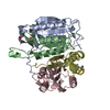
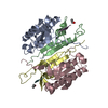
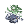
| Title | Crystal Structure of Caspase-7 with Inhibitor AC-WEHD-CHO | ||||||
 Components Components |
| ||||||
 Keywords Keywords | HYDROLASE/HYDROLASE INHIBITOR / CYSTEINE PROTEASE / APOPTOSIS / THIOL PROTEASE / ZYMOGEN / HYDROLASE-HYDROLASE INHIBITOR COMPLEX | ||||||
| Function / homology |  Function and homology information Function and homology informationlymphocyte apoptotic process / caspase-7 / positive regulation of plasma membrane repair / cellular response to staurosporine / SMAC, XIAP-regulated apoptotic response / Activation of caspases through apoptosome-mediated cleavage / plasma membrane repair / SMAC (DIABLO) binds to IAPs / SMAC(DIABLO)-mediated dissociation of IAP:caspase complexes / fibroblast apoptotic process ...lymphocyte apoptotic process / caspase-7 / positive regulation of plasma membrane repair / cellular response to staurosporine / SMAC, XIAP-regulated apoptotic response / Activation of caspases through apoptosome-mediated cleavage / plasma membrane repair / SMAC (DIABLO) binds to IAPs / SMAC(DIABLO)-mediated dissociation of IAP:caspase complexes / fibroblast apoptotic process / ceramide biosynthetic process / execution phase of apoptosis / Apoptotic cleavage of cellular proteins / Caspase-mediated cleavage of cytoskeletal proteins / response to UV / striated muscle cell differentiation / cysteine-type peptidase activity / protein catabolic process / protein maturation / protein processing / fibrillar center / peptidase activity / positive regulation of neuron apoptotic process / heart development / cellular response to lipopolysaccharide / neuron apoptotic process / aspartic-type endopeptidase activity / defense response to bacterium / cysteine-type endopeptidase activity / apoptotic process / proteolysis / : / RNA binding / nucleoplasm / nucleus / plasma membrane / cytoplasm / cytosol Similarity search - Function | ||||||
| Biological species |  Homo sapiens (human) Homo sapiens (human) | ||||||
| Method |  X-RAY DIFFRACTION / SYNCHROTRON / MOLECULAR REPLACEMENT / Resolution: 2.6 Å X-RAY DIFFRACTION / SYNCHROTRON / MOLECULAR REPLACEMENT / Resolution: 2.6 Å | ||||||
 Authors Authors | Agniswamy, J. / Fang, B. / Weber, I. | ||||||
 Citation Citation | Journal: Febs J. / Year: 2007 Title: Plasticity of S2-S4 specificity pockets of executioner caspase-7 revealed by structural and kinetic analysis. Authors: Agniswamy, J. / Fang, B. / Weber, I.T. | ||||||
| History |
|
- Structure visualization
| Structure viewer | Molecule: MolmilJmol/JSmol |
|---|
- Downloads & links
-Download
| PDBx/mmCIF format | 2qlj.cif.gz | 111.8 KB | Display | PDBx/mmCIF format |
|---|---|---|---|---|
| PDB format | pdb2qlj.ent.gz | 85.1 KB | Display | PDB format |
| PDBx/mmJSON format | 2qlj.json.gz | Tree view | PDBx/mmJSON format | |
| Others |  Other downloads Other downloads |
-Validation report
| Arichive directory | https://data.pdbj.org/pub/pdb/validation_reports/ql/2qljftp://data.pdbj.org/pub/pdb/validation_reports/ql/2qlj | HTTPS FTP |
|---|
-Related structure data
| Related structure data |  2ql5C  2ql7C  2ql9C 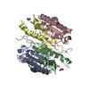 2qlbC 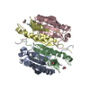 2qlfC 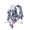 1f1jS C: citing same article ( S: Starting model for refinement |
|---|---|
| Similar structure data |


-Links
 PDBj
PDBj


- Assembly
| Deposited unit | 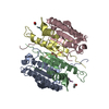
| ||||||||
|---|---|---|---|---|---|---|---|---|---|
| 1 |
| ||||||||
| Unit cell |
|
-Components
-Protein , 2 types, 4 molecules ACBD
| #1: Protein | Mass: 19564.598 Da / Num. of mol.: 2 / Fragment: P20 subunit Source method: isolated from a genetically manipulated source Source: (gene. exp.) Homo sapiens (human) / Gene: CASP7, MCH3 / Plasmid: PET23b / Species (production host): Escherichia coli / Production host:  #2: Protein | Mass: 11352.862 Da / Num. of mol.: 2 / Fragment: P10 subunit Source method: isolated from a genetically manipulated source Source: (gene. exp.) Homo sapiens (human) / Gene: CASP7, MCH3 / Plasmid: PET23b / Species (production host): Escherichia coli / Production host: |
|---|
-Protein/peptide , 2 types, 3 molecules EFG
| #3: Protein/peptide |   Type: Peptide-like / Class: Inhibitor / Mass: 598.627 Da / Num. of mol.: 2 / Source method: obtained synthetically Type: Peptide-like / Class: Inhibitor / Mass: 598.627 Da / Num. of mol.: 2 / Source method: obtained syntheticallyReferences: N-acetyl-L-tryptophyl-L-alpha-glutamyl-N-[(1R)-2-carboxy-1-formylethyl]-3-(1H-imidazol-3-ium-4-yl)-L-alaninamide #4: Protein/peptide | | Mass: 527.508 Da / Num. of mol.: 1 / Source method: obtained synthetically |
|---|
-Non-polymers , 2 types, 53 molecules 

| #5: Chemical | ChemComp-CIT /  Mass: 192.124 Da / Num. of mol.: 1 / Source method: obtained synthetically / Formula: C6H8O7 Mass: 192.124 Da / Num. of mol.: 1 / Source method: obtained synthetically / Formula: C6H8O7 |
|---|---|
| #6: Water | ChemComp-HOH / Mass: 18.015 Da / Num. of mol.: 52 / Source method: isolated from a natural source / Formula: H2O |
-Details
| Compound details | THE INHIBITOR IS COVALENTLY |
|---|
-Experimental details
-Experiment
| Experiment | Method: X-RAY DIFFRACTION / Number of used crystals: 1 |
|---|
- Sample preparation
| Crystal | Density Matthews: 3.34 Å3/Da / Density % sol: 63.12 % |
|---|---|
| Crystal grow | Temperature: 298 K / Method: vapor diffusion, hanging drop / pH: 6.7 Details: 14.5% PEG 3350, 0.3M diammonium hydrogen citrate, 10mM DTT, pH 6.7, VAPOR DIFFUSION, HANGING DROP, temperature 298K |
-Data collection
| Diffraction | Mean temperature: 100 K |
|---|---|
| Diffraction source | Source: SYNCHROTRON / Site: APS  / Beamline: 22-ID / Wavelength: 1 Å / Beamline: 22-ID / Wavelength: 1 Å |
| Detector | Type: MARMOSAIC 300 mm CCD / Detector: CCD / Date: Mar 27, 2007 |
| Radiation | Monochromator: si 220 / Protocol: SINGLE WAVELENGTH / Monochromatic (M) / Laue (L): M / Scattering type: x-ray |
| Radiation wavelength | Wavelength: 1 Å / Relative weight: 1 |
| Reflection | Resolution: 2.6→50 Å / Num. all: 24929 / Num. obs: 21440 / % possible obs: 86 % / Observed criterion σ(F): 2 / Observed criterion σ(I): 2 / Redundancy: 3.4 % / Rmerge(I) obs: 0.067 / Net I/σ(I): 16.73 |
| Reflection shell | Resolution: 2.6→2.69 Å / Redundancy: 3 % / Rmerge(I) obs: 0.348 / Mean I/σ(I) obs: 3.25 / Num. unique all: 1620 / % possible all: 60.7 |
- Processing
| Software |
| |||||||||||||||||||||||||
|---|---|---|---|---|---|---|---|---|---|---|---|---|---|---|---|---|---|---|---|---|---|---|---|---|---|---|
| Refinement | Method to determine structure: MOLECULAR REPLACEMENT Starting model: PDB entry 1F1J Resolution: 2.6→50 Å / Cross valid method: THROUGHOUT / σ(F): 0 / σ(I): 0 / Stereochemistry target values: Engh & Huber
| |||||||||||||||||||||||||
| Refinement step | Cycle: LAST / Resolution: 2.6→50 Å
|
