[English] 日本語
Yorodumi
- PDB-2qf7: Crystal structure of a complete multifunctional pyruvate carboxyl... -

+
Open data


ID or keywords:

Loading...

-
Basic information

Entry
Database: PDB / ID: 2qf7
TitleCrystal structure of a complete multifunctional pyruvate carboxylase from Rhizobium etli
ComponentsPyruvate carboxylase protein
KeywordsLIGASE / MULTI-DOMAIN / MULTI-FUNCTIONAL / BIOTIN-DEPENDENT
Function / homology
Function and homology information


pyruvate carboxylase / pyruvate carboxylase activity / pyruvate metabolic process / gluconeogenesis / ATP binding / metal ion binding
Similarity search - Function
pyruvate carboxylase f1077a mutant fold / pyruvate carboxylase f1077a mutant domain / Conserved carboxylase domain / Pyruvate carboxylase / Carboxylase, conserved domain / Conserved carboxylase domain / Pyruvate carboxyltransferase / HMGL-like / Pyruvate carboxyltransferase domain. / Biotin-binding site ...pyruvate carboxylase f1077a mutant fold / pyruvate carboxylase f1077a mutant domain / Conserved carboxylase domain / Pyruvate carboxylase / Carboxylase, conserved domain / Conserved carboxylase domain / Pyruvate carboxyltransferase / HMGL-like / Pyruvate carboxyltransferase domain. / Biotin-binding site / Biotin-requiring enzymes attachment site. / Biotin carboxylase-like, N-terminal domain / Biotin carboxylase, C-terminal / Biotin carboxylation domain / Biotin carboxylase, N-terminal domain / Biotin carboxylase C-terminal domain / Biotin carboxylation domain profile. / Biotin carboxylase C-terminal domain / Rossmann fold - #20 / Carbamoyl-phosphate synthase subdomain signature 1. / Carbamoyl-phosphate synthetase large subunit-like, ATP-binding domain / Carbamoyl-phosphate synthase L chain, ATP binding domain / Biotin-requiring enzyme / RNA polymerase II/Efflux pump adaptor protein, barrel-sandwich hybrid domain / Biotinyl/lipoyl domain profile. / Biotin/lipoyl attachment / Rudiment single hybrid motif / Single hybrid motif / Cyclin A; domain 1 / ATP-grasp fold, B domain / Pre-ATP-grasp domain superfamily / ATP-grasp fold / ATP-grasp fold profile. / D-amino Acid Aminotransferase; Chain A, domain 1 / Homeodomain-like / Aldolase class I / OB fold (Dihydrolipoamide Acetyltransferase, E2P) / Carbamoyl-phosphate synthase subdomain signature 2. / Aldolase-type TIM barrel / Arc Repressor Mutant, subunit A / TIM Barrel / Roll / Alpha-Beta Barrel / Beta Barrel / Rossmann fold / 2-Layer Sandwich / Orthogonal Bundle / 3-Layer(aba) Sandwich / Mainly Beta / Mainly Alpha / Alpha Beta
Similarity search - Domain/homology
PHOSPHOTHIOPHOSPHORIC ACID-ADENYLATE ESTER / COENZYME A / FORMIC ACID / Pyruvate carboxylase
Similarity search - Component
Biological speciesRhizobium etli (bacteria)
MethodX-RAY DIFFRACTION / SYNCHROTRON / SAD / Resolution: 2 Å
AuthorsSt Maurice, M. / Surinya, K.H. / Rayment, I.
CitationJournal: Science / Year: 2007
Title: Domain architecture of pyruvate carboxylase, a biotin-dependent multifunctional enzyme
Authors: St Maurice, M. / Reinhardt, L. / Surinya, K.H. / Attwood, P.V. / Wallace, J.C. / Cleland, W.W. / Rayment, I.
History
DepositionJun 27, 2007Deposition site: RCSB / Processing site: RCSB
Revision 1.0Sep 4, 2007Provider: repository / Type: Initial release
Revision 1.1Jul 13, 2011Group: Advisory / Derived calculations ...Advisory / Derived calculations / Source and taxonomy / Version format compliance
Revision 1.2Feb 15, 2012Group: Non-polymer description
Remark 600 HETEROGEN The distance between atoms PG and S1G in ligand AGS is 1.95 angstroms.

-
Structure visualization

Structure viewerMolecule:
MolmilJmol/JSmol

Downloads & links

-
Assembly

Deposited unit
A: Pyruvate carboxylase protein
B: Pyruvate carboxylase protein
hetero molecules


Theoretical massNumber of molelcules
Total (without water)257,54120
Polymers255,1242
Non-polymers2,41718
Water23,7441318
1
A: Pyruvate carboxylase protein
B: Pyruvate carboxylase protein
hetero molecules

A: Pyruvate carboxylase protein
B: Pyruvate carboxylase protein
hetero molecules


Theoretical massNumber of molelcules
Total (without water)515,08140
Polymers510,2484
Non-polymers4,83336
Water724
TypeNameSymmetry operationNumber
identity operation1_555x,y,z1
crystal symmetry operation2_655-x+1,y,-z1
Buried area25700 Å2
ΔGint-411 kcal/mol
Surface area147890 Å2
MethodPISA, PQS
Unit cell
Length a, b, c (Å)234.729, 93.258, 137.222
Angle α, β, γ (deg.)90.000, 107.330, 90.000
Int Tables number5
Space group name H-MC121
Components on special symmetry positions
IDModelComponents
11A-1548-

HOH

Details
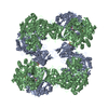
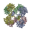
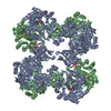
The biological unit is a tetramer. The following symmetry operation is required to build the tetramer from the dimer in the asymmetric unit: rotation matrix: -1 0 0 0 1 0 0 0 -1 translation vector: 234.72900 0.00000 0.00000 The equivalent Eulerean rotation is Phi = 0.00000, Theta = 180.00000, Psi = 0.00000

-
Components

-
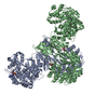
Protein , 1 types, 2 molecules AB

#1: Protein Pyruvate carboxylase protein /


Mass: 127562.039 Da / Num. of mol.: 2
Source method: isolated from a genetically manipulated source
Source: (gene. exp.) Rhizobium etli (bacteria) / Strain: CFN 42 / Gene: pyc / Plasmid: pET17b / Production host: Escherichia coli (E. coli) / Strain (production host): Rosetta(DE3) / References: UniProt: Q2K340, pyruvate carboxylase

-
Non-polymers , 8 types, 1336 molecules

#2: Chemical
ChemComp-MG / MAGNESIUM ION


Mass: 24.305 Da / Num. of mol.: 6 / Source method: obtained synthetically / Formula: Mg
#3: Chemical ChemComp-ZN / ZINC ION


Mass: 65.409 Da / Num. of mol.: 2 / Source method: obtained synthetically / Formula: Zn
#4: Chemical
ChemComp-CL / CHLORIDE ION / Chloride


Mass: 35.453 Da / Num. of mol.: 4 / Source method: obtained synthetically / Formula: Cl
#5: Chemical ChemComp-COA / COENZYME A / Coenzyme A


Mass: 767.534 Da / Num. of mol.: 1 / Source method: obtained synthetically / Formula: C21H36N7O16P3S
#6: Chemical ChemComp-AGS / PHOSPHOTHIOPHOSPHORIC ACID-ADENYLATE ESTER / ATP-GAMMA-S / ADENOSINE 5'-(3-THIOTRIPHOSPHATE) / ADENOSINE 5'-(GAMMA-THIOTRIPHOSPHATE) / ADENOSINE-5'-DIPHOSPHATE MONOTHIOPHOSPHATE


Mass: 523.247 Da / Num. of mol.: 2 / Source method: obtained synthetically / Formula: C10H16N5O12P3S / Comment: ATP-gamma-S, energy-carrying molecule analogue*YM
#7: Chemical ChemComp-FMT / FORMIC ACID / Formic acid


Mass: 46.025 Da / Num. of mol.: 2 / Source method: obtained synthetically / Formula: CH2O2
#8: Chemical ChemComp-GOL / GLYCEROL / GLYCERIN / PROPANE-1,2,3-TRIOL / Glycerol


Mass: 92.094 Da / Num. of mol.: 1 / Source method: obtained synthetically / Formula: C3H8O3
#9: Water ChemComp-HOH / water / Water


Mass: 18.015 Da / Num. of mol.: 1318 / Source method: isolated from a natural source / Formula: H2O

-
Experimental details

-
Experiment

ExperimentMethod: X-RAY DIFFRACTION / Number of used crystals: 1

-
Sample preparation

Crystal
IDDensity Matthews3/Da)Density % sol (%)
12.8456.7
2
Crystal grow
Temperature (K)Crystal-IDMethodpHDetails
2981evaporation5.67.5 mg/mL PC, 2.5 mM ATP-gamma-S, 1 mM ethyl CoA, 280 mM sodium formate, 70 mM sodium acetate, 60 mM calcium chloride, 5.9% PEG 8K, pH 5.6, EVAPORATION, temperature 298K
2982vapor diffusion, hanging drop5.6400 mM sodium formate, 80 mM sodium acetate, 80 mM calcium chloride, 14 mM 3-(N,N-Dimethyltetradecylammonio)propanesulfonate, 9% PEG 8K, pH 5.6, VAPOR DIFFUSION, HANGING DROP, temperature 298K

-
Data collection

Diffraction sourceSource: SYNCHROTRON / Site: APS / Beamline: 19-BM / Wavelength: 0.97907 Å
DetectorType: SBC-3 / Detector: CCD / Date: Aug 18, 2006
RadiationProtocol: SINGLE WAVELENGTH / Monochromatic (M) / Laue (L): M / Scattering type: x-ray
Radiation wavelengthWavelength: 0.97907 Å / Relative weight: 1
ReflectionResolution: 2→30 Å / Num. all: 184567 / Num. obs: 184296 / % possible obs: 96.7 % / Observed criterion σ(F): 0 / Observed criterion σ(I): 0 / Redundancy: 4 % / Biso Wilson estimate: 25.8 Å2 / Rsym value: 0.057 / Net I/σ(I): 20.1
Reflection shellResolution: 2→2.07 Å / Redundancy: 3.3 % / Mean I/σ(I) obs: 3.1 / Rsym value: 0.321 / % possible all: 86

-
Phasing

PhasingMethod: SAD
Phasing MAD set site
IDAtom type symbolB isoFract xFract yFract zOccupancy
1Se29.2230.6930.0030.4531.359
2Se6.2070.8670.2670.1310.681
3Se40.9880.7120.3710.4051.396
4Se26.6720.2430.0140.4471.045
5Se20.7680.6840.4440.4681.233
6Se19.2770.1770.3910.3160.879
7Se36.6910.9690.0230.4441.183
8Se40.4030.1770.4640.4561.174
9Se37.8560.1870.1820.451.342
10Se32.8710.5870.4310.2271.176
11Se43.9080.5890.3860.2521.252
12Se7.7180.8640.2670.1410.712
13Se22.5720.1560.3620.3910.901
14Se39.2140.1450.1240.4561.1
15Se48.7210.6690.010.4350.755
16Se35.0810.1110.0320.2891.059
17Se40.3550.6150.0230.2261.316
18Se54.0350.5980.3840.2191.614
19Se35.5970.1270.4390.1960.857
20Se45.0350.8520.0310.181.134
21Se48.0750.0710.4810.2131.279
22Se600.1850.4860.2221.646
23Se600.3210.3630.0511.463
24Se32.7890.6940.3840.3750.726
25Se44.4680.6640.4830.3331.287
26Se45.2570.0880.2830.240.894
27Se600.4320.470.0361.255
28Se59.5140.650.0250.451.101
29Se35.2650.1870.4760.2590.754
30Se41.8170.1820.0860.4880.815
31Se35.7660.6540.3730.4570.707
32Se51.4760.630.4380.4980.848
33Se600.7850.3920.0310.992
34Se42.0330.7510.3130.2070.663
35Se30.5110.1010.30.1870.493
36Se44.4860.5650.3860.2530.677
37Se23.4060.6280.3730.3010.389
38Se39.6140.1190.2050.2130.523
39Se37.7860.2930.020.1370.408
40Se36.5240.1650.0320.3780.461
41Se8.0310.6730.010.3650.251
42Se600.9610.460.2581.032
43Se32.9580.2160.1190.0670.289
44Se600.1760.340.1680.477
45Se600.0210.1110.2660.637
46Se600.9990.0420.30.918
47Se25.9580.1510.3480.2810.269
48Se37.0310.3970.3060.1130.243
49Se600.3890.2660.1090.378
50Se10.7120.0180.4620.075
51Se600.2440.1790.210.871
Phasing dmFOM : 0.68 / FOM acentric: 0.68 / FOM centric: 0.66 / Reflection: 75150 / Reflection acentric: 72619 / Reflection centric: 2531
Phasing dm shell
Resolution (Å)FOM FOM acentricFOM centricReflectionReflection acentricReflection centric
7.5-42.2170.940.940.8635473204343
4.7-7.50.880.880.761098910403586
3.8-4.70.860.870.741354613063483
3.3-3.80.760.760.651333912942397
2.8-3.30.560.560.472180321297506
2.6-2.80.380.380.331192611710216

-
Processing

Software
NameVersionClassificationNB
SOLVE2.1phasing
RESOLVE2.1phasing
REFMAC5.2.0005refinement
PDB_EXTRACT2data extraction
HKL-2000data collection
DENZOdata reduction
SCALEPACKdata scaling
RefinementMethod to determine structure: SAD / Resolution: 2→131.31 Å / Cor.coef. Fo:Fc: 0.957 / Cor.coef. Fo:Fc free: 0.934 / SU B: 6.093 / SU ML: 0.095 / TLS residual ADP flag: LIKELY RESIDUAL / Cross valid method: THROUGHOUT / σ(F): 0 / ESU R: 0.15 / ESU R Free: 0.146 / Stereochemistry target values: MAXIMUM LIKELIHOOD / Details: HYDROGENS HAVE BEEN ADDED IN THE RIDING POSITIONS
RfactorNum. reflection% reflectionSelection details
Rfree0.223 9277 5 %RANDOM
Rwork0.178 ---
obs0.18 184296 96.92 %-
Solvent computationIon probe radii: 0.8 Å / Shrinkage radii: 0.8 Å / VDW probe radii: 1.2 Å / Solvent model: MASK
Displacement parametersBiso mean: 25.599 Å2
Baniso -1Baniso -2Baniso -3
1--0.99 Å20 Å2-0.86 Å2
2---0.7 Å20 Å2
3---1.18 Å2
Refine analyzeLuzzati coordinate error obs: 0.322 Å
Refinement stepCycle: LAST / Resolution: 2→131.31 Å
ProteinNucleic acidLigandSolventTotal
Num. atoms15782 0 118 1318 17218
Refine LS restraints
Refine-IDTypeDev idealDev ideal targetNumber
X-RAY DIFFRACTIONr_bond_refined_d0.0160.02216682
X-RAY DIFFRACTIONr_angle_refined_deg1.6231.97222780
X-RAY DIFFRACTIONr_dihedral_angle_1_deg6.76152206
X-RAY DIFFRACTIONr_dihedral_angle_2_deg33.46624.008716
X-RAY DIFFRACTIONr_dihedral_angle_3_deg14.836152664
X-RAY DIFFRACTIONr_dihedral_angle_4_deg18.30615118
X-RAY DIFFRACTIONr_chiral_restr0.1210.22575
X-RAY DIFFRACTIONr_gen_planes_refined0.0070.0212791
X-RAY DIFFRACTIONr_nbd_refined0.2120.28016
X-RAY DIFFRACTIONr_nbtor_refined0.3080.211549
X-RAY DIFFRACTIONr_xyhbond_nbd_refined0.1570.21286
X-RAY DIFFRACTIONr_metal_ion_refined0.1980.25
X-RAY DIFFRACTIONr_symmetry_vdw_refined0.2010.294
X-RAY DIFFRACTIONr_symmetry_hbond_refined0.1770.242
X-RAY DIFFRACTIONr_mcbond_it0.8931.510947
X-RAY DIFFRACTIONr_mcangle_it1.351217093
X-RAY DIFFRACTIONr_scbond_it2.20536545
X-RAY DIFFRACTIONr_scangle_it3.1874.55638
LS refinement shellResolution: 2→2.054 Å / Total num. of bins used: 20
RfactorNum. reflection% reflection
Rfree0.252 591 -
Rwork0.195 11771 -
obs-12362 88.57 %
Refinement TLS params.

Details: PH?A@?=?e ?jM=O>^)><,?v??\>3>< ?>P>+>16?#y> / Method: refined / Refine-ID: X-RAY DIFFRACTION

IDL112)L122)L132)L222)L232)L332)S11 (Å °)S12 (Å °)S13 (Å °)S21 (Å °)S22 (Å °)S23 (Å °)S31 (Å °)S32 (Å °)S33 (Å °)T112)T122)T132)T222)T232)T332)Origin x (Å)Origin y (Å)Origin z (Å)
11.9903-0.12610.03330.78430.11850.53570.0056-0.08950.26530.0283-0.06350.1524-0.1968-0.0980.0579-0.0787-0.011-0.0043-0.0342-0.0356-0.0882126.4151100.965351.7892
23.5796-0.42040.40892.1995-0.58023.4096-0.0368-0.02710.04310.01630.01050.0823-0.1005-0.25340.0263-0.1225-0.01840.03320.05120.0004-0.1779132.91492.57129.365
30.76160.05690.17480.8265-0.1041.1608-0.03770.07580.1576-0.0454-0.01750.0045-0.1374-0.040.0553-0.1507-0.0135-0.0107-0.20690.0268-0.1198160.007104.2973-14.0319
413.84321.68680.46714.74870.5975.5976-0.2410.02020.2958-0.12690.17580.0907-0.2534-0.01920.06530.1525-0.01930.02680.196-0.01830.1227107.8754108.204422.0584
51.6618-0.25470.0480.7891-0.29571.14720.03010.0725-0.3023-0.0708-0.0736-0.03250.26220.15240.0434-0.09730.00820.019-0.03660.0355-0.1093149.688575.667242.9189
63.79160.58561.31464.3094-0.34052.00090.1279-0.0891-0.24090.0859-0.0788-0.38690.15960.3082-0.0491-0.0471-0.0171-0.07670.03040.03140.0803110.184769.579638.6018
70.5280.05760.44231.18560.13931.05850.0761-0.1036-0.1160.16840.01590.01060.0488-0.0569-0.092-0.127-0.02370.0219-0.13910.0264-0.128873.583565.457229.6981
Refinement TLS group
IDRefine-IDRefine TLS-IDAuth asym-IDLabel asym-IDAuth seq-IDLabel seq-ID
1X-RAY DIFFRACTION1AA1 - 45512 - 466
2X-RAY DIFFRACTION2AA471 - 489482 - 500
3X-RAY DIFFRACTION2AA1014 - 10731025 - 1084
4X-RAY DIFFRACTION3AA525 - 1000536 - 1011
5X-RAY DIFFRACTION4AA1079 - 11521090 - 1163
6X-RAY DIFFRACTION5BB1 - 45512 - 466
7X-RAY DIFFRACTION6BB471 - 489482 - 500
8X-RAY DIFFRACTION6BB1014 - 10681025 - 1079
9X-RAY DIFFRACTION7BB525 - 1000536 - 1011

+
About Yorodumi

-
News

-
Feb 9, 2022. New format data for meta-information of EMDB entries

New format data for meta-information of EMDB entries

  • Version 3 of the EMDB header file is now the official format.
  • The previous official version 1.9 will be removed from the archive.

Related info.:EMDB header

External links:wwPDB to switch to version 3 of the EMDB data model

-
Aug 12, 2020. Covid-19 info

Covid-19 info

URL: https://pdbj.org/emnavi/covid19.php

New page: Covid-19 featured information page in EM Navigator.

Related info.:Covid-19 info / Mar 5, 2020. Novel coronavirus structure data

+
Mar 5, 2020. Novel coronavirus structure data

Novel coronavirus structure data

Related info.:Yorodumi Speices / Aug 12, 2020. Covid-19 info

External links:COVID-19 featured content - PDBj / Molecule of the Month (242):Coronavirus Proteases

+
Jan 31, 2019. EMDB accession codes are about to change! (news from PDBe EMDB page)

EMDB accession codes are about to change! (news from PDBe EMDB page)

  • The allocation of 4 digits for EMDB accession codes will soon come to an end. Whilst these codes will remain in use, new EMDB accession codes will include an additional digit and will expand incrementally as the available range of codes is exhausted. The current 4-digit format prefixed with “EMD-” (i.e. EMD-XXXX) will advance to a 5-digit format (i.e. EMD-XXXXX), and so on. It is currently estimated that the 4-digit codes will be depleted around Spring 2019, at which point the 5-digit format will come into force.
  • The EM Navigator/Yorodumi systems omit the EMD- prefix.

Related info.:Q: What is EMD? / ID/Accession-code notation in Yorodumi/EM Navigator

External links:EMDB Accession Codes are Changing Soon! / Contact to PDBj

+
Jul 12, 2017. Major update of PDB

Major update of PDB

  • wwPDB released updated PDB data conforming to the new PDBx/mmCIF dictionary.
  • This is a major update changing the version number from 4 to 5, and with Remediation, in which all the entries are updated.
  • In this update, many items about electron microscopy experimental information are reorganized (e.g. em_software).
  • Now, EM Navigator and Yorodumi are based on the updated data.

External links:wwPDB Remediation / Enriched Model Files Conforming to OneDep Data Standards Now Available in the PDB FTP Archive

-
Yorodumi

Thousand views of thousand structures

  • Yorodumi is a browser for structure data from EMDB, PDB, SASBDB, etc.
  • This page is also the successor to EM Navigator detail page, and also detail information page/front-end page for Omokage search.
  • The word "yorodu" (or yorozu) is an old Japanese word meaning "ten thousand". "mi" (miru) is to see.

Related info.:EMDB / PDB / SASBDB / Comparison of 3 databanks / Yorodumi Search / Aug 31, 2016. New EM Navigator & Yorodumi / Yorodumi Papers / Jmol/JSmol / Function and homology information / Changes in new EM Navigator and Yorodumi

Read more