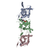
The biological assembly is a hexamer, but hexamer is divided by crystallographic symmetry, trimer occupies the asymmetric unit. Trimer model was generated from Sept2 (2QA5, submitted recently).
-
Components
#1: Protein
Septin-2 / Protein NEDD5
Mass: 41550.508 Da / Num. of mol.: 1 Source method: isolated from a genetically manipulated source Source: (gene. exp.) Homo sapiens (human) / Gene: SEPT2, DIFF6, KIAA0158, NEDD5 / Production host: Escherichia coli (E. coli) / Strain (production host): Rosetta BL21 / References: UniProt: Q15019
#2: Protein
Septin-6
Mass: 48948.723 Da / Num. of mol.: 1 Source method: isolated from a genetically manipulated source Source: (gene. exp.) Homo sapiens (human) / Gene: SEPT6, KIAA0128, SEP2 / Production host: Escherichia coli (E. coli) / References: UniProt: Q14141
#3: Protein
Septin-7 / CDC10 protein homolog
Mass: 48837.945 Da / Num. of mol.: 1 Source method: isolated from a genetically manipulated source Source: (gene. exp.) Homo sapiens (human) / Gene: SEPT7, CDC10 / Production host: Escherichia coli (E. coli) / References: UniProt: Q16181
Monochromator: Si 111 CHANNEL / Protocol: SINGLE WAVELENGTH / Monochromatic (M) / Laue (L): M / Scattering type: x-ray
Radiation wavelength
Wavelength: 1.07188 Å / Relative weight: 1
Reflection
Resolution: 4→50 Å / Num. obs: 42485 / Redundancy: 16.2 % / Rsym value: 0.229 / Net I/σ(I): 11.42
Reflection shell
Resolution: 4→4.1 Å / Rmerge(I) obs: 0.379 / Mean I/σ(I) obs: 3.98 / % possible all: 98.9
-
Processing
Software
Name
Version
Classification
NB
REFMAC
5.2.0005
refinement
PDB_EXTRACT
2
dataextraction
SOLVE
phasing
Refinement
Method to determine structure: MIRAS / Resolution: 4→49.15 Å / Cor.coef. Fo:Fc: 0.796 / Cor.coef. Fo:Fc free: 0.787 / SU B: 82.738 / SU ML: 0.616 / Cross valid method: THROUGHOUT / ESU R: 0.7 / ESU R Free: 0.54 Stereochemistry target values: MAXIMUM LIKELIHOOD WITH PHASES Details: HYDROGENS HAVE BEEN ADDED IN THE RIDING POSITIONS. Sept6 (Chain B) and Sept7 (Chain C) will have poly-alanine stretches due low resolution.
Rfactor
Num. reflection
% reflection
Selection details
Rfree
0.39208
2125
5 %
RANDOM
Rwork
0.37554
-
-
-
obs
0.37637
40358
98.27 %
-
Solvent computation
Ion probe radii: 0.8 Å / Shrinkage radii: 0.8 Å / VDW probe radii: 1.2 Å / Solvent model: MASK
Displacement parameters
Biso mean: 78.174 Å2
Baniso -1
Baniso -2
Baniso -3
1-
-14.93 Å2
0 Å2
0 Å2
2-
-
-14.93 Å2
0 Å2
3-
-
-
29.85 Å2
Refinement step
Cycle: LAST / Resolution: 4→49.15 Å
Protein
Nucleic acid
Ligand
Solvent
Total
Num. atoms
4407
0
88
0
4495
Refine LS restraints
Refine-ID
Type
Dev ideal
Dev ideal target
Number
X-RAY DIFFRACTION
r_bond_refined_d
0.057
0.022
4612
X-RAY DIFFRACTION
r_bond_other_d
X-RAY DIFFRACTION
r_angle_refined_deg
4.719
1.954
6311
X-RAY DIFFRACTION
r_angle_other_deg
X-RAY DIFFRACTION
r_dihedral_angle_1_deg
18.875
5
681
X-RAY DIFFRACTION
r_dihedral_angle_2_deg
51.524
25.258
97
X-RAY DIFFRACTION
r_dihedral_angle_3_deg
30.803
15
461
X-RAY DIFFRACTION
r_dihedral_angle_4_deg
5.216
15
6
X-RAY DIFFRACTION
r_chiral_restr
0.247
0.2
808
X-RAY DIFFRACTION
r_gen_planes_refined
0.014
0.02
3366
X-RAY DIFFRACTION
r_gen_planes_other
X-RAY DIFFRACTION
r_nbd_refined
0.475
0.2
3293
X-RAY DIFFRACTION
r_nbd_other
X-RAY DIFFRACTION
r_nbtor_refined
0.375
0.2
2772
X-RAY DIFFRACTION
r_nbtor_other
X-RAY DIFFRACTION
r_xyhbond_nbd_refined
0.403
0.2
320
X-RAY DIFFRACTION
r_xyhbond_nbd_other
X-RAY DIFFRACTION
r_metal_ion_refined
X-RAY DIFFRACTION
r_metal_ion_other
X-RAY DIFFRACTION
r_symmetry_vdw_refined
0.339
0.2
15
X-RAY DIFFRACTION
r_symmetry_vdw_other
X-RAY DIFFRACTION
r_symmetry_hbond_refined
X-RAY DIFFRACTION
r_symmetry_hbond_other
X-RAY DIFFRACTION
r_symmetry_metal_ion_refined
X-RAY DIFFRACTION
r_symmetry_metal_ion_other
X-RAY DIFFRACTION
r_mcbond_it
1.747
1.5
3815
X-RAY DIFFRACTION
r_mcbond_other
X-RAY DIFFRACTION
r_mcangle_it
2.844
2
5299
X-RAY DIFFRACTION
r_scbond_it
3.276
3
1223
X-RAY DIFFRACTION
r_scangle_it
5.123
4.5
1012
X-RAY DIFFRACTION
r_rigid_bond_restr
X-RAY DIFFRACTION
r_sphericity_free
X-RAY DIFFRACTION
r_sphericity_bonded
LS refinement shell
Resolution: 4→4.103 Å / Total num. of bins used: 20
Rfactor
Num. reflection
% reflection
Rfree
0.454
156
-
Rwork
0.467
2948
-
obs
-
-
98.95 %
+
About Yorodumi
-
News
-
Feb 9, 2022. New format data for meta-information of EMDB entries
New format data for meta-information of EMDB entries
Version 3 of the EMDB header file is now the official format.
The previous official version 1.9 will be removed from the archive.
In the structure databanks used in Yorodumi, some data are registered as the other names, "COVID-19 virus" and "2019-nCoV". Here are the details of the virus and the list of structure data.
Jan 31, 2019. EMDB accession codes are about to change! (news from PDBe EMDB page)
EMDB accession codes are about to change! (news from PDBe EMDB page)
The allocation of 4 digits for EMDB accession codes will soon come to an end. Whilst these codes will remain in use, new EMDB accession codes will include an additional digit and will expand incrementally as the available range of codes is exhausted. The current 4-digit format prefixed with “EMD-” (i.e. EMD-XXXX) will advance to a 5-digit format (i.e. EMD-XXXXX), and so on. It is currently estimated that the 4-digit codes will be depleted around Spring 2019, at which point the 5-digit format will come into force.
The EM Navigator/Yorodumi systems omit the EMD- prefix.
Related info.:Q: What is EMD? / ID/Accession-code notation in Yorodumi/EM Navigator
Yorodumi is a browser for structure data from EMDB, PDB, SASBDB, etc.
This page is also the successor to EM Navigator detail page, and also detail information page/front-end page for Omokage search.
The word "yorodu" (or yorozu) is an old Japanese word meaning "ten thousand". "mi" (miru) is to see.
Related info.:EMDB / PDB / SASBDB / Comparison of 3 databanks / Yorodumi Search / Aug 31, 2016. New EM Navigator & Yorodumi / Yorodumi Papers / Jmol/JSmol / Function and homology information / Changes in new EM Navigator and Yorodumi
 Movie
Movie Controller


 Open data
 Basic information
 Components
 Keywords
 Function and homology information
 Homo sapiens (human)
 X-RAY DIFFRACTION /
  Authors
 Citation
 Structure visualization
 Downloads & links
 Other downloads
 PDBj
PDBj Assembly


 Type: RNA linking / Mass: 443.201 Da / Num. of mol.: 2 / Source method: obtained synthetically / Formula: C10H15N5O11P2 / Comment: GDP, energy-carrying molecule*YM
 Mass: 523.180 Da / Num. of mol.: 1 / Source method: obtained synthetically / Formula: C10H16N5O14P3 / Comment: GTP, energy-carrying molecule*YM
 Sample preparation
 / Beamline: X10SA / Wavelength: 1.07188 Å
 Processing