- PDB-2q81: Crystal Structure of the Miz-1 BTB/POZ domain -
+
Open data
ID or keywords:
Loading...
-
Basic information
Entry
Database: PDB / ID: 2q81
Title
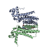
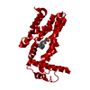
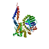
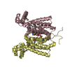
Crystal Structure of the Miz-1 BTB/POZ domain
Components
Miz-1 protein
Keywords
TRANSCRIPTION / BTB/POZ domain
Function / homology
Function and homology information
ectoderm development / XBP1(S) activates chaperone genes / gastrulation with mouth forming second / regulation of immune system process / G1 to G0 transition / negative regulation of cell cycle / core promoter sequence-specific DNA binding / regulation of cytokine production / protein-DNA complex / DNA-binding transcription repressor activity, RNA polymerase II-specific ...ectoderm development / XBP1(S) activates chaperone genes / gastrulation with mouth forming second / regulation of immune system process / G1 to G0 transition / negative regulation of cell cycle / core promoter sequence-specific DNA binding / regulation of cytokine production / protein-DNA complex / DNA-binding transcription repressor activity, RNA polymerase II-specific / transcription coactivator binding / DNA-binding transcription activator activity, RNA polymerase II-specific / DNA-binding transcription factor binding / RNA polymerase II cis-regulatory region sequence-specific DNA binding / DNA-binding transcription factor activity / negative regulation of cell population proliferation / negative regulation of transcription by RNA polymerase II / positive regulation of transcription by RNA polymerase II / protein-containing complex / zinc ion binding / nucleoplasm Similarity search - Function
C2H2-type zinc finger / Potassium Channel Kv1.1; Chain A / Potassium Channel Kv1.1; Chain A / BTB/POZ domain / BTB domain profile. / Zinc finger, C2H2 type / Broad-Complex, Tramtrack and Bric a brac / BTB/POZ domain / zinc finger / Zinc finger C2H2 type domain profile. ...C2H2-type zinc finger / Potassium Channel Kv1.1; Chain A / Potassium Channel Kv1.1; Chain A / BTB/POZ domain / BTB domain profile. / Zinc finger, C2H2 type / Broad-Complex, Tramtrack and Bric a brac / BTB/POZ domain / zinc finger / Zinc finger C2H2 type domain profile. / Zinc finger C2H2 superfamily / Zinc finger C2H2 type domain signature. / SKP1/BTB/POZ domain superfamily / Zinc finger C2H2-type / 2-Layer Sandwich / Alpha Beta Similarity search - Domain/homology
Miz-1protein / Zinc finger and BTB domain-containing protein 17 / Zinc finger protein 151 / Myc-interacting zinc ...Zinc finger and BTB domain-containing protein 17 / Zinc finger protein 151 / Myc-interacting zinc finger protein
Mass: 13071.004 Da / Num. of mol.: 4 / Fragment: BTB domain, residues 2-115 Source method: isolated from a genetically manipulated source Source: (gene. exp.) Homo sapiens (human) / Gene: ZBTB17, MIZ1, ZNF151 / Plasmid: pGEX6P1 / Production host: Escherichia coli (E. coli) / Strain (production host): BL21(DE3)pLysS / References: UniProt: Q13105
In the structure databanks used in Yorodumi, some data are registered as the other names, "COVID-19 virus" and "2019-nCoV". Here are the details of the virus and the list of structure data.
Jan 31, 2019. EMDB accession codes are about to change! (news from PDBe EMDB page)
EMDB accession codes are about to change! (news from PDBe EMDB page)
The allocation of 4 digits for EMDB accession codes will soon come to an end. Whilst these codes will remain in use, new EMDB accession codes will include an additional digit and will expand incrementally as the available range of codes is exhausted. The current 4-digit format prefixed with “EMD-” (i.e. EMD-XXXX) will advance to a 5-digit format (i.e. EMD-XXXXX), and so on. It is currently estimated that the 4-digit codes will be depleted around Spring 2019, at which point the 5-digit format will come into force.
The EM Navigator/Yorodumi systems omit the EMD- prefix.
Related info.:Q: What is EMD? / ID/Accession-code notation in Yorodumi/EM Navigator
Yorodumi is a browser for structure data from EMDB, PDB, SASBDB, etc.
This page is also the successor to EM Navigator detail page, and also detail information page/front-end page for Omokage search.
The word "yorodu" (or yorozu) is an old Japanese word meaning "ten thousand". "mi" (miru) is to see.
Related info.:EMDB / PDB / SASBDB / Comparison of 3 databanks / Yorodumi Search / Aug 31, 2016. New EM Navigator & Yorodumi / Yorodumi Papers / Jmol/JSmol / Function and homology information / Changes in new EM Navigator and Yorodumi
 Movie
Movie Controller


 Open data
 Basic information
 Components
 Keywords
 Function and homology information
 Homo sapiens (human)
 X-RAY DIFFRACTION /
  Authors
 Citation
 Structure visualization
 Downloads & links
 Other downloads


 PDBj
PDBj


 Assembly
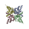


 Mass: 194.226 Da / Num. of mol.: 1 / Source method: obtained synthetically / Formula: C8H18O5 / Comment: precipitant*YM
 Mass: 18.015 Da / Num. of mol.: 239 / Source method: isolated from a natural source / Formula: H2O
 Sample preparation
 / Beamline: PX14.2 / Wavelength: 0.979 Å
 Processing