 Movie
Movie Controller

[English] 日本語
 Yorodumi
- PDB-2pzz: 2.2 A resolution crystal structure of UPF0201 protein from Methan... -
+ Open data

- Basic information
| Entry | Database: PDB / ID: 2pzz | ||||||
|---|---|---|---|---|---|---|---|
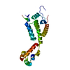
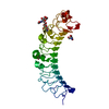
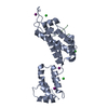
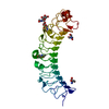
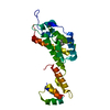
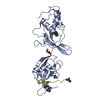
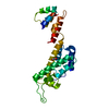
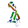
| Title | 2.2 A resolution crystal structure of UPF0201 protein from Methanococcus jannaschii | ||||||
 Components Components | UPF0201 protein MJ1564 | ||||||
 Keywords Keywords | STRUCTURAL GENOMICS / UNKNOWN FUNCTION / UPF0201 / MJ1564 / 10077a3 / Methanococcus jannaschii / PSI2 / NYSGXRC / Protein Structure Initiative / New York SGX Research Center for Structural Genomics | ||||||
| Function / homology | Uncharacterised protein family UPF0201 / RNA binding / 50s Ribosomal Protein L5; Chain: A, / Ribosomal protein L5 / Ribosomal protein L5 domain superfamily / 2-Layer Sandwich / Alpha Beta / UPF0201 protein MJ1564 Function and homology information Function and homology information | ||||||
| Biological species |   Methanocaldococcus jannaschii DSM 2661 (archaea) Methanocaldococcus jannaschii DSM 2661 (archaea) | ||||||
| Method |  X-RAY DIFFRACTION / SYNCHROTRON / SAD / Resolution: 2.2 Å X-RAY DIFFRACTION / SYNCHROTRON / SAD / Resolution: 2.2 Å | ||||||
 Authors Authors | Rao, K.N. / Burley, S.K. / Swaminathan, S. / New York SGX Research Center for Structural Genomics (NYSGXRC) | ||||||
 Citation Citation | Journal: Plos One / Year: 2008 Title: UPF201 archaeal specific family members reveal structural similarity to RNA-binding proteins but low likelihood for RNA-binding function. Authors: Rao, K.N. / Burley, S.K. / Swaminathan, S. | ||||||
| History |
|
- Structure visualization
| Structure viewer | Molecule: MolmilJmol/JSmol |
|---|
- Downloads & links
-Download
| PDBx/mmCIF format | 2pzz.cif.gz | 107.7 KB | Display | PDBx/mmCIF format |
|---|---|---|---|---|
| PDB format | pdb2pzz.ent.gz | 84.4 KB | Display | PDB format |
| PDBx/mmJSON format | 2pzz.json.gz | Tree view | PDBx/mmJSON format | |
| Others |  Other downloads Other downloads |
-Validation report
| Summary document | 2pzz_validation.pdf.gz | 456.5 KB | Display | wwPDB validaton report |
|---|---|---|---|---|
| Full document | 2pzz_full_validation.pdf.gz | 470.3 KB | Display | |
| Data in XML | 2pzz_validation.xml.gz | 21 KB | Display | |
| Data in CIF | 2pzz_validation.cif.gz | 28.4 KB | Display | |
| Arichive directory | https://data.pdbj.org/pub/pdb/validation_reports/pz/2pzzftp://data.pdbj.org/pub/pdb/validation_reports/pz/2pzz | HTTPS FTP |
-Related structure data
| Related structure data | 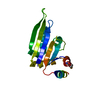 2nrqC  2nwuC 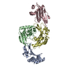 2ogkC C: citing same article ( |
|---|---|
| Similar structure data | |
| Other databases |


-Links
 PDBj
PDBj- Assembly
| Deposited unit | 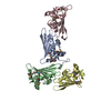
| ||||||||
|---|---|---|---|---|---|---|---|---|---|
| 1 | 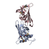
| ||||||||
| 2 | 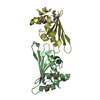
| ||||||||
| Unit cell |
|
-Components
| #1: Protein | Mass: 16896.023 Da / Num. of mol.: 4 Source method: isolated from a genetically manipulated source Source: (gene. exp.) Methanocaldococcus jannaschii DSM 2661 (archaea)Species: Methanocaldococcus jannaschii / Strain: DSM 2661, JAL-1, JCM 10045, NBRC 100440 / Gene: MJ1564 / Plasmid: pSGX3(BC) / Species (production host): Escherichia coli / Production host:  #2: Water | ChemComp-HOH / |  Mass: 18.015 Da / Num. of mol.: 83 / Source method: isolated from a natural source / Formula: H2O Mass: 18.015 Da / Num. of mol.: 83 / Source method: isolated from a natural source / Formula: H2OHas protein modification | Y | |
|---|
-Experimental details
-Experiment
| Experiment | Method: X-RAY DIFFRACTION / Number of used crystals: 2 |
|---|
- Sample preparation
| Crystal | Density Matthews: 2.29 Å3/Da / Density % sol: 46.28 % |
|---|---|
| Crystal grow | Temperature: 293 K / Method: vapor diffusion, sitting drop / pH: 7.5 Details: Ammonium acetate, HEPES Buffer, PEG3350, pH 7.5, VAPOR DIFFUSION, SITTING DROP, temperature 293K |
-Data collection
| Diffraction | Mean temperature: 100 K |
|---|---|
| Diffraction source | Source: SYNCHROTRON / Site: NSLS  / Beamline: X12C / Wavelength: 0.9793 Å / Beamline: X12C / Wavelength: 0.9793 Å |
| Detector | Type: ADSC QUANTUM 210 / Detector: CCD / Date: May 1, 2007 / Details: Mirrors |
| Radiation | Monochromator: Si III / Protocol: SINGLE WAVELENGTH / Monochromatic (M) / Laue (L): M / Scattering type: x-ray |
| Radiation wavelength | Wavelength: 0.9793 Å / Relative weight: 1 |
| Reflection | Resolution: 2.2→50 Å / Num. all: 30128 / Num. obs: 30128 / % possible obs: 98.9 % / Observed criterion σ(F): 0 / Observed criterion σ(I): 0 / Redundancy: 7.2 % / Biso Wilson estimate: 25.7 Å2 / Rmerge(I) obs: 0.07 / Net I/σ(I): 14.6 |
| Reflection shell | Resolution: 2.2→2.28 Å / Redundancy: 5.3 % / Rmerge(I) obs: 0.278 / Mean I/σ(I) obs: 2.4 / Num. unique all: 2775 / % possible all: 90.9 |
- Processing
| Software |
| ||||||||||||||||||||||||||||||||||||
|---|---|---|---|---|---|---|---|---|---|---|---|---|---|---|---|---|---|---|---|---|---|---|---|---|---|---|---|---|---|---|---|---|---|---|---|---|---|
| Refinement | Method to determine structure: SAD / Resolution: 2.2→47.23 Å / Rfactor Rfree error: 0.01 / Data cutoff high absF: 120713.55 / Data cutoff low absF: 0 / Isotropic thermal model: RESTRAINED / Cross valid method: THROUGHOUT / σ(F): 0 / Stereochemistry target values: Engh & Huber Details: Residues listed as missing in Remark 465 are due to lack of electron density. Residues with missing atoms listed in Remark 470 are due to lack of electron density for side chains and modeled as alanines.
| ||||||||||||||||||||||||||||||||||||
| Solvent computation | Solvent model: FLAT MODEL / Bsol: 45.4452 Å2 / ksol: 0.364126 e/Å3 | ||||||||||||||||||||||||||||||||||||
| Displacement parameters | Biso mean: 46.4 Å2
| ||||||||||||||||||||||||||||||||||||
| Refine analyze |
| ||||||||||||||||||||||||||||||||||||
| Refinement step | Cycle: LAST / Resolution: 2.2→47.23 Å
| ||||||||||||||||||||||||||||||||||||
| Refine LS restraints |
| ||||||||||||||||||||||||||||||||||||
| LS refinement shell | Resolution: 2.2→2.34 Å / Rfactor Rfree error: 0.028 / Total num. of bins used: 6
| ||||||||||||||||||||||||||||||||||||
| Xplor file |
|
