 Movie
Movie Controller


+ Open data

- Basic information
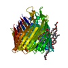
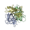
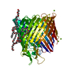
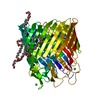
| Entry | Database: PDB / ID: 2pzh | ||||||
|---|---|---|---|---|---|---|---|
| Title | YbgC thioesterase (Hp0496) from Helicobacter pylori | ||||||
 Components Components | Hypothetical protein HP_0496 | ||||||
 Keywords Keywords | HYDROLASE / lipid / acyl-CoA / bacterial membrane / Tol-Pal system / thioesterase / Hot-dog fold | ||||||
| Function / homology |  Function and homology information Function and homology informationfatty acyl-CoA hydrolase activity / Hydrolases; Acting on ester bonds; Thioester hydrolases / lipid metabolic process Similarity search - Function | ||||||
| Biological species |   Helicobacter pylori (bacteria) Helicobacter pylori (bacteria) | ||||||
| Method |  X-RAY DIFFRACTION / SYNCHROTRON / MOLECULAR REPLACEMENT / Resolution: 1.7 Å X-RAY DIFFRACTION / SYNCHROTRON / MOLECULAR REPLACEMENT / Resolution: 1.7 Å | ||||||
 Authors Authors | Angelini, A. / Cendron, L. / Goncalves, S. / Zanotti, G. / Terradot, L. | ||||||
 Citation Citation | Journal: Proteins / Year: 2008 Title: Structural and enzymatic characterization of HP0496, a YbgC thioesterase from Helicobacter pylori. Authors: Angelini, A. / Cendron, L. / Goncalves, S. / Zanotti, G. / Terradot, L. #1: Journal: BMC Bioinformatics / Year: 2004Title: The Hotdog fold: wrapping up a superfamily of thioesterases and dehydratases Authors: Dillon, S.C. / Bateman, A. #2: Journal: J.Biol.Chem. / Year: 1998Title: The three-dimensional structure of 4-hydroxybenzoyl-CoA thioesterase from Pseudomonas sp. Strain CBS-3 Authors: Benning, M.M. / Wesemberg, G. / Liu, R. / Taylor, K.L. / Dunway-Mariano, D. / Holden, H.M. | ||||||
| History |
|
- Structure visualization
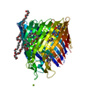
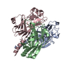
| Structure viewer | Molecule: MolmilJmol/JSmol |
|---|
- Downloads & links
-Download
| PDBx/mmCIF format | 2pzh.cif.gz | 124.8 KB | Display | PDBx/mmCIF format |
|---|---|---|---|---|
| PDB format | pdb2pzh.ent.gz | 98.2 KB | Display | PDB format |
| PDBx/mmJSON format | 2pzh.json.gz | Tree view | PDBx/mmJSON format | |
| Others |  Other downloads Other downloads |
-Validation report
| Arichive directory | https://data.pdbj.org/pub/pdb/validation_reports/pz/2pzhftp://data.pdbj.org/pub/pdb/validation_reports/pz/2pzh | HTTPS FTP |
|---|
-Related structure data
| Related structure data |  1s5uS S: Starting model for refinement |
|---|---|
| Similar structure data |

-Links
 PDBj
PDBj
- Assembly
| Deposited unit | 
| ||||||||||||||||||||||||||||||
|---|---|---|---|---|---|---|---|---|---|---|---|---|---|---|---|---|---|---|---|---|---|---|---|---|---|---|---|---|---|---|---|
| 1 |
| ||||||||||||||||||||||||||||||
| Unit cell |
| ||||||||||||||||||||||||||||||
| Noncrystallographic symmetry (NCS) | NCS domain:
NCS domain segments: Component-ID: 1 / Ens-ID: 1 / Beg auth comp-ID: MET / Beg label comp-ID: MET / End auth comp-ID: ALA / End label comp-ID: ALA / Refine code: 5 / Auth seq-ID: 1 - 132 / Label seq-ID: 3 - 134
| ||||||||||||||||||||||||||||||
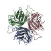
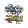
| Details | The biological assembly is the homo-tetramer (chains A,B,C,D) |
-Components
| #1: Protein | Mass: 15854.352 Da / Num. of mol.: 4 Source method: isolated from a genetically manipulated source Source: (gene. exp.) Helicobacter pylori (bacteria) / Strain: 26695 / Gene: HP0496 / Plasmid: pET151/D-TOPO / Production host: #2: Water | ChemComp-HOH / |  Mass: 18.015 Da / Num. of mol.: 295 / Source method: isolated from a natural source / Formula: H2O Mass: 18.015 Da / Num. of mol.: 295 / Source method: isolated from a natural source / Formula: H2O |
|---|
-Experimental details
-Experiment
| Experiment | Method: X-RAY DIFFRACTION / Number of used crystals: 1 |
|---|
- Sample preparation
| Crystal | Density Matthews: 2.09 Å3/Da / Density % sol: 41.18 % |
|---|---|
| Crystal grow | Temperature: 298 K / Method: vapor diffusion, hanging drop / pH: 8.5 Details: 100mM Tris, 20% Ethanol, pH 8.5, VAPOR DIFFUSION, HANGING DROP, temperature 298K |
-Data collection
| Diffraction | Mean temperature: 100 K |
|---|---|
| Diffraction source | Source: SYNCHROTRON / Site: ESRF  / Beamline: ID14-3 / Wavelength: 0.931 / Beamline: ID14-3 / Wavelength: 0.931 |
| Detector | Type: ADSC QUANTUM 4 / Detector: CCD / Date: Jun 19, 2006 |
| Radiation | Protocol: SINGLE WAVELENGTH / Monochromatic (M) / Laue (L): M / Scattering type: x-ray |
| Radiation wavelength | Wavelength: 0.931 Å / Relative weight: 1 |
| Reflection | Resolution: 1.7→45 Å / Num. all: 53355 / Num. obs: 53355 / % possible obs: 90.3 % / Observed criterion σ(F): 0 / Observed criterion σ(I): 0 / Redundancy: 4.8 % / Rmerge(I) obs: 0.05 / Net I/σ(I): 22 |
| Reflection shell | Resolution: 1.7→1.79 Å / Redundancy: 4.2 % / Rmerge(I) obs: 0.5 / Mean I/σ(I) obs: 2.7 / Num. unique all: 6452 / % possible all: 76.5 |
- Processing
| Software |
| ||||||||||||||||||||||||||||||||||||||||||||||||||||||||||||||||||||||||||||||||||||||||||||||||||||||||||||||||||||||||||||||||||||||||||||||||||||||||||||||||||||||||||
|---|---|---|---|---|---|---|---|---|---|---|---|---|---|---|---|---|---|---|---|---|---|---|---|---|---|---|---|---|---|---|---|---|---|---|---|---|---|---|---|---|---|---|---|---|---|---|---|---|---|---|---|---|---|---|---|---|---|---|---|---|---|---|---|---|---|---|---|---|---|---|---|---|---|---|---|---|---|---|---|---|---|---|---|---|---|---|---|---|---|---|---|---|---|---|---|---|---|---|---|---|---|---|---|---|---|---|---|---|---|---|---|---|---|---|---|---|---|---|---|---|---|---|---|---|---|---|---|---|---|---|---|---|---|---|---|---|---|---|---|---|---|---|---|---|---|---|---|---|---|---|---|---|---|---|---|---|---|---|---|---|---|---|---|---|---|---|---|---|---|---|---|
| Refinement | Method to determine structure: MOLECULAR REPLACEMENT Starting model: 1S5U Resolution: 1.7→45 Å / Cor.coef. Fo:Fc: 0.943 / Cor.coef. Fo:Fc free: 0.924 / SU B: 2.542 / SU ML: 0.086 / Cross valid method: THROUGHOUT / σ(F): 0 / σ(I): 0.5 / ESU R: 0.145 / ESU R Free: 0.138 / Stereochemistry target values: MAXIMUM LIKELIHOOD / Details: HYDROGENS HAVE BEEN ADDED IN THE RIDING POSITIONS
| ||||||||||||||||||||||||||||||||||||||||||||||||||||||||||||||||||||||||||||||||||||||||||||||||||||||||||||||||||||||||||||||||||||||||||||||||||||||||||||||||||||||||||
| Solvent computation | Ion probe radii: 0.8 Å / Shrinkage radii: 0.8 Å / VDW probe radii: 1.4 Å / Solvent model: MASK | ||||||||||||||||||||||||||||||||||||||||||||||||||||||||||||||||||||||||||||||||||||||||||||||||||||||||||||||||||||||||||||||||||||||||||||||||||||||||||||||||||||||||||
| Displacement parameters | Biso mean: 20.851 Å2
| ||||||||||||||||||||||||||||||||||||||||||||||||||||||||||||||||||||||||||||||||||||||||||||||||||||||||||||||||||||||||||||||||||||||||||||||||||||||||||||||||||||||||||
| Refinement step | Cycle: LAST / Resolution: 1.7→45 Å
| ||||||||||||||||||||||||||||||||||||||||||||||||||||||||||||||||||||||||||||||||||||||||||||||||||||||||||||||||||||||||||||||||||||||||||||||||||||||||||||||||||||||||||
| Refine LS restraints |
| ||||||||||||||||||||||||||||||||||||||||||||||||||||||||||||||||||||||||||||||||||||||||||||||||||||||||||||||||||||||||||||||||||||||||||||||||||||||||||||||||||||||||||
| Refine LS restraints NCS | Ens-ID: 1 / Refine-ID: X-RAY DIFFRACTION
| ||||||||||||||||||||||||||||||||||||||||||||||||||||||||||||||||||||||||||||||||||||||||||||||||||||||||||||||||||||||||||||||||||||||||||||||||||||||||||||||||||||||||||
| LS refinement shell | Resolution: 1.7→1.744 Å / Total num. of bins used: 20
|
