 ムービー
ムービー コントローラー


+ データを開く

- 基本情報
基本情報
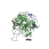
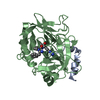
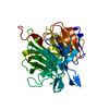
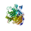
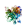
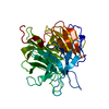
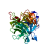
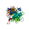
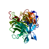
| 登録情報 | データベース: PDB / ID: 2pux | ||||||
|---|---|---|---|---|---|---|---|
| タイトル | Crystal structure of murine thrombin in complex with the extracellular fragment of murine PAR3 | ||||||
 要素 要素 |
| ||||||
 キーワード キーワード | HYDROLASE / SERINE PROTEASE | ||||||
| 機能・相同性 |  機能・相同性情報 機能・相同性情報proteinase-activated receptor activity / Common Pathway of Fibrin Clot Formation / Platelet Aggregation (Plug Formation) / Gamma-carboxylation of protein precursors / Transport of gamma-carboxylated protein precursors from the endoplasmic reticulum to the Golgi apparatus / Intrinsic Pathway of Fibrin Clot Formation / Removal of aminoterminal propeptides from gamma-carboxylated proteins / thrombin-activated receptor signaling pathway / thrombin-activated receptor activity / Peptide ligand-binding receptors ...proteinase-activated receptor activity / Common Pathway of Fibrin Clot Formation / Platelet Aggregation (Plug Formation) / Gamma-carboxylation of protein precursors / Transport of gamma-carboxylated protein precursors from the endoplasmic reticulum to the Golgi apparatus / Intrinsic Pathway of Fibrin Clot Formation / Removal of aminoterminal propeptides from gamma-carboxylated proteins / thrombin-activated receptor signaling pathway / thrombin-activated receptor activity / Peptide ligand-binding receptors / Thrombin signalling through proteinase activated receptors (PARs) / Regulation of Complement cascade / G alpha (q) signalling events / Cell surface interactions at the vascular wall / positive regulation of Rho protein signal transduction / cytolysis by host of symbiont cells / positive regulation of phospholipase C-activating G protein-coupled receptor signaling pathway / thrombospondin receptor activity / thrombin / neutrophil-mediated killing of gram-negative bacterium / ligand-gated ion channel signaling pathway / negative regulation of astrocyte differentiation / positive regulation of collagen biosynthetic process / negative regulation of cytokine production involved in inflammatory response / positive regulation of blood coagulation / regulation of cytosolic calcium ion concentration / fibrinolysis / positive regulation of release of sequestered calcium ion into cytosol / acute-phase response / G protein-coupled receptor activity / negative regulation of proteolysis / lipopolysaccharide binding / positive regulation of insulin secretion / platelet activation / positive regulation of protein localization to nucleus / positive regulation of reactive oxygen species metabolic process / blood coagulation / antimicrobial humoral immune response mediated by antimicrobial peptide / heparin binding / peptidase activity / regulation of cell shape / phospholipase C-activating G protein-coupled receptor signaling pathway / positive regulation of cell growth / regulation of gene expression / collagen-containing extracellular matrix / endopeptidase activity / positive regulation of phosphatidylinositol 3-kinase/protein kinase B signal transduction / cell surface receptor signaling pathway / receptor ligand activity / positive regulation of protein phosphorylation / G protein-coupled receptor signaling pathway / apical plasma membrane / external side of plasma membrane / serine-type endopeptidase activity / signaling receptor binding / calcium ion binding / positive regulation of cell population proliferation / protein-containing complex / proteolysis / extracellular space / plasma membrane 類似検索 - 分子機能 | ||||||
| 生物種 |  | ||||||
| 手法 |  X線回折 / シンクロトロン / 分子置換 / 解像度: 2 Å X線回折 / シンクロトロン / 分子置換 / 解像度: 2 Å | ||||||
 データ登録者 データ登録者 | Bah, A. / Chen, Z. / Bush-Pelc, L.A. / Mathews, F.S. / Di Cera, E. | ||||||
 引用 引用 | ジャーナル: Proc.Natl.Acad.Sci.Usa / 年: 2007 タイトル: Crystal structures of murine thrombin in complex with the extracellular fragments of murine protease-activated receptors PAR3 and PAR4. 著者: Bah, A. / Chen, Z. / Bush-Pelc, L.A. / Mathews, F.S. / Di Cera, E. #1: ジャーナル: J.Biol.Chem. / 年: 2004タイトル: Molecular Dissection of Na+ Binding to Thrombin. 著者: Pineda, A.O. / Carrell, C.J. / Bush, L.A. / Prasad, S. / Caccia, S. / Chen, Z. / Mathews, F.S. / Di Cera, E. #2: ジャーナル: J.Biol.Chem. / 年: 2007タイトル: Structural Basis of Na+ Activation mimicry in murine. 著者: Marino, F. / Chen, Z. / Ergenekan, C.E. / Bush, L.A. / Mathews, F.S. / Di Cera, E. | ||||||
| 履歴 |
|
- 構造の表示
構造の表示
| 構造ビューア | 分子: MolmilJmol/JSmol |
|---|
- ダウンロードとリンク
-ダウンロード
| PDBx/mmCIF形式 | 2pux.cif.gz | 85.4 KB | 表示 | PDBx/mmCIF形式 |
|---|---|---|---|---|
| PDB形式 | pdb2pux.ent.gz | 62.9 KB | 表示 | PDB形式 |
| PDBx/mmJSON形式 | 2pux.json.gz | ツリー表示 | PDBx/mmJSON形式 | |
| その他 |  その他のダウンロード その他のダウンロード |
-検証レポート
| 文書・要旨 | 2pux_validation.pdf.gz | 464.6 KB | 表示 | wwPDB検証レポート |
|---|---|---|---|---|
| 文書・詳細版 | 2pux_full_validation.pdf.gz | 471.7 KB | 表示 | |
| XML形式データ | 2pux_validation.xml.gz | 18.1 KB | 表示 | |
| CIF形式データ | 2pux_validation.cif.gz | 26.3 KB | 表示 | |
| アーカイブディレクトリ | https://data.pdbj.org/pub/pdb/validation_reports/pu/2puxftp://data.pdbj.org/pub/pdb/validation_reports/pu/2pux | HTTPS FTP |
-関連構造データ


-リンク
 PDBj
PDBj


- 集合体
集合体
| 登録構造単位 | 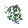
| ||||||||
|---|---|---|---|---|---|---|---|---|---|
| 1 |
| ||||||||
| 単位格子 |
| ||||||||
| 詳細 | The biological assembly is a monomer. |
-要素
| #1: タンパク質・ペプチド | 分子量: 5105.731 Da / 分子数: 1 / 由来タイプ: 組換発現 / 由来: (組換発現) 発現宿主:  Cricetulus griseus (モンゴルキヌゲネズミ) Cricetulus griseus (モンゴルキヌゲネズミ)参照: UniProt: P19221 |
|---|---|
| #2: タンパク質 | 分子量: 29952.625 Da / 分子数: 1 / 由来タイプ: 組換発現 / 由来: (組換発現) 発現宿主: Cricetulus griseus (モンゴルキヌゲネズミ)参照: UniProt: P19221 |
| #3: タンパク質・ペプチド | 分子量: 1568.636 Da / 分子数: 1 / 由来タイプ: 合成 / 詳細: MIDWEST Biotech Inc. / 参照: UniProt: O08675 |
| #4: 糖 | ChemComp-NAG /   タイプ: D-saccharide, beta linking / 分子量: 221.208 Da / 分子数: 1 / 由来タイプ: 組換発現 / 式: C8H15NO6 タイプ: D-saccharide, beta linking / 分子量: 221.208 Da / 分子数: 1 / 由来タイプ: 組換発現 / 式: C8H15NO6 |
| #5: 水 | ChemComp-HOH /  分子量: 18.015 Da / 分子数: 295 / 由来タイプ: 天然 / 式: H2O 分子量: 18.015 Da / 分子数: 295 / 由来タイプ: 天然 / 式: H2O |
| Has protein modification | Y |
-実験情報
-実験
| 実験 | 手法: X線回折 / 使用した結晶の数: 1 |
|---|
- 試料調製
試料調製
| 結晶 | マシュー密度: 2.86 Å3/Da / 溶媒含有率: 56.99 % |
|---|---|
| 結晶化 | 温度: 295 K / 手法: 蒸気拡散法, ハンギングドロップ法 / pH: 7.5 詳細: 20% PEG 10000, 100 mM HEPES, pH 7.5, VAPOR DIFFUSION, HANGING DROP, temperature 295K |
-データ収集
| 回折 | 平均測定温度: 100 K |
|---|---|
| 放射光源 | 由来: シンクロトロン / サイト: APS  / ビームライン: 14-BM-C / 波長: 0.9 Å / ビームライン: 14-BM-C / 波長: 0.9 Å |
| 検出器 | タイプ: ADSC QUANTUM 315 / 検出器: CCD / 日付: 2007年4月6日 |
| 放射 | プロトコル: SINGLE WAVELENGTH / 単色(M)・ラウエ(L): M / 散乱光タイプ: x-ray |
| 放射波長 | 波長: 0.9 Å / 相対比: 1 |
| 反射 | 解像度: 2→40 Å / Num. all: 28405 / Num. obs: 27610 / % possible obs: 97.2 % / Observed criterion σ(F): -1 / Observed criterion σ(I): -1 / 冗長度: 3.5 % / Biso Wilson estimate: 5.5 Å2 / Rmerge(I) obs: 0.1 / Net I/σ(I): 10.6 |
| 反射 シェル | 解像度: 2→2.07 Å / 冗長度: 2.8 % / Rmerge(I) obs: 0.306 / Mean I/σ(I) obs: 2.9 / Num. unique all: 2506 / % possible all: 88.6 |
- 解析
解析
| ソフトウェア |
| ||||||||||||||||||||||||||||||||||||
|---|---|---|---|---|---|---|---|---|---|---|---|---|---|---|---|---|---|---|---|---|---|---|---|---|---|---|---|---|---|---|---|---|---|---|---|---|---|
| 精密化 | 構造決定の手法: 分子置換 開始モデル: PDB entry 1SHH 解像度: 2→27.08 Å / Rfactor Rfree error: 0.006 / Data cutoff high absF: 187825.98 / Data cutoff low absF: 0 / Isotropic thermal model: RESTRAINED / 交差検証法: THROUGHOUT / σ(F): 0 / 立体化学のターゲット値: Engh & Huber
| ||||||||||||||||||||||||||||||||||||
| 原子変位パラメータ | Biso mean: 21.9 Å2 | ||||||||||||||||||||||||||||||||||||
| Refine analyze |
| ||||||||||||||||||||||||||||||||||||
| 精密化ステップ | サイクル: LAST / 解像度: 2→27.08 Å
| ||||||||||||||||||||||||||||||||||||
| 拘束条件 |
| ||||||||||||||||||||||||||||||||||||
| LS精密化 シェル | 解像度: 2→2.13 Å / Rfactor Rfree error: 0.022 / Total num. of bins used: 6
|
