 Movie
Movie Controller

[English] 日本語
 Yorodumi
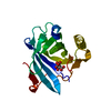
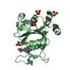
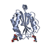
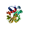
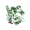
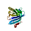
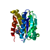
- PDB-2prb: crystal structure of aminoglycoside acetyltransferase AAC(6')-Ib ... -
+ Open data

- Basic information
| Entry | Database: PDB / ID: 2prb | ||||||
|---|---|---|---|---|---|---|---|
| Title | crystal structure of aminoglycoside acetyltransferase AAC(6')-Ib in complex whith coenzyme A | ||||||
 Components Components | Aminoglycoside 6-N-acetyltransferase type Ib11 | ||||||
 Keywords Keywords | TRANSFERASE / GNAT / aminoglycoside acetyltransferase | ||||||
| Function / homology |  Function and homology information Function and homology informationaminoglycoside 6'-N-acetyltransferase / aminoglycoside 6'-N-acetyltransferase activity / response to antibiotic Similarity search - Function | ||||||
| Biological species |  Salmonella typhimurium (bacteria) Salmonella typhimurium (bacteria) | ||||||
| Method |  X-RAY DIFFRACTION / SYNCHROTRON / MOLECULAR REPLACEMENT / Resolution: 1.8 Å X-RAY DIFFRACTION / SYNCHROTRON / MOLECULAR REPLACEMENT / Resolution: 1.8 Å | ||||||
 Authors Authors | Maurice, F. / Broutin, I. / Podglajen, I. / Benas, P. / Collatz, E. / Dardel, F. | ||||||
 Citation Citation | Journal: Embo Rep. / Year: 2008 Title: Enzyme structural plasticity and the emergence of broad-spectrum antibiotic resistance. Authors: Maurice, F. / Broutin, I. / Podglajen, I. / Benas, P. / Collatz, E. / Dardel, F. | ||||||
| History |
|
- Structure visualization
| Structure viewer | Molecule: MolmilJmol/JSmol |
|---|
- Downloads & links
-Download
| PDBx/mmCIF format | 2prb.cif.gz | 53.9 KB | Display | PDBx/mmCIF format |
|---|---|---|---|---|
| PDB format | pdb2prb.ent.gz | 36.6 KB | Display | PDB format |
| PDBx/mmJSON format | 2prb.json.gz | Tree view | PDBx/mmJSON format | |
| Others |  Other downloads Other downloads |
-Validation report
| Arichive directory | https://data.pdbj.org/pub/pdb/validation_reports/pr/2prbftp://data.pdbj.org/pub/pdb/validation_reports/pr/2prb | HTTPS FTP |
|---|
-Related structure data
| Related structure data |  2pr8SC 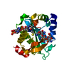 2qirC S: Starting model for refinement C: citing same article ( |
|---|---|
| Similar structure data |


-Links
 PDBj
PDBj


- Assembly
| Deposited unit | 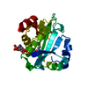
| ||||||||
|---|---|---|---|---|---|---|---|---|---|
| 1 |
| ||||||||
| Unit cell |
|
-Components
| #1: Protein | Mass: 21921.439 Da / Num. of mol.: 1 / Mutation: L106Q,S107L Source method: isolated from a genetically manipulated source Source: (gene. exp.) Salmonella typhimurium (bacteria) / Plasmid: pET101 / Production host: References: UniProt: Q8GLI5, aminoglycoside 6'-N-acetyltransferase |
|---|---|
| #2: Chemical | ChemComp-COA /   Mass: 767.534 Da / Num. of mol.: 1 / Source method: obtained synthetically / Formula: C21H36N7O16P3S Mass: 767.534 Da / Num. of mol.: 1 / Source method: obtained synthetically / Formula: C21H36N7O16P3S |
| #3: Water | ChemComp-HOH /  Mass: 18.015 Da / Num. of mol.: 172 / Source method: isolated from a natural source / Formula: H2O Mass: 18.015 Da / Num. of mol.: 172 / Source method: isolated from a natural source / Formula: H2O |
-Experimental details
-Experiment
| Experiment | Method: X-RAY DIFFRACTION / Number of used crystals: 1 |
|---|
- Sample preparation
| Crystal | Density Matthews: 2.78 Å3/Da / Density % sol: 55.68 % |
|---|---|
| Crystal grow | Temperature: 291 K / Method: vapor diffusion, hanging drop / pH: 7 Details: 1.5 M K2HPO4 0.06M NaH2PO4 0.1M guanidine-HCl, pH 7, VAPOR DIFFUSION, HANGING DROP, temperature 291K |
-Data collection
| Diffraction | Mean temperature: 77 K |
|---|---|
| Diffraction source | Source: SYNCHROTRON / Site: ESRF  / Beamline: ID14-3 / Wavelength: 0.931 Å / Beamline: ID14-3 / Wavelength: 0.931 Å |
| Detector | Type: ADSC QUANTUM 4r / Detector: CCD / Date: Mar 3, 2007 |
| Radiation | Monochromator: diamond 111 / Protocol: SINGLE WAVELENGTH / Monochromatic (M) / Laue (L): M / Scattering type: x-ray |
| Radiation wavelength | Wavelength: 0.931 Å / Relative weight: 1 |
| Reflection | Resolution: 1.8→53.63 Å / Num. all: 23842 / Num. obs: 23836 / % possible obs: 100 % / Observed criterion σ(F): 0 / Observed criterion σ(I): 1 / Redundancy: 13.5 % / Rmerge(I) obs: 0.059 / Rsym value: 0.059 / Net I/σ(I): 8.3 |
| Reflection shell | Resolution: 1.8→1.9 Å / Redundancy: 10.5 % / Rmerge(I) obs: 0.183 / Mean I/σ(I) obs: 4.1 / Num. measured all: 35626 / Num. unique all: 3394 / Rsym value: 0.183 / % possible all: 100 |
-Phasing
| Phasing MR |
|
|---|
- Processing
| Software |
| ||||||||||||||||||||||||||||||||||||||||||||||||||||||||||||||||||||||||||||||||||||||||||
|---|---|---|---|---|---|---|---|---|---|---|---|---|---|---|---|---|---|---|---|---|---|---|---|---|---|---|---|---|---|---|---|---|---|---|---|---|---|---|---|---|---|---|---|---|---|---|---|---|---|---|---|---|---|---|---|---|---|---|---|---|---|---|---|---|---|---|---|---|---|---|---|---|---|---|---|---|---|---|---|---|---|---|---|---|---|---|---|---|---|---|---|
| Refinement | Method to determine structure: MOLECULAR REPLACEMENT Starting model: pdb entry 2PR8 Resolution: 1.8→40.76 Å / Cor.coef. Fo:Fc: 0.936 / Cor.coef. Fo:Fc free: 0.909 / SU B: 2.2 / SU ML: 0.071 / Cross valid method: THROUGHOUT / σ(F): 0 / ESU R: 0.12 / ESU R Free: 0.11 / Stereochemistry target values: MAXIMUM LIKELIHOOD / Details: HYDROGENS HAVE BEEN ADDED IN THE RIDING POSITIONS
| ||||||||||||||||||||||||||||||||||||||||||||||||||||||||||||||||||||||||||||||||||||||||||
| Solvent computation | Ion probe radii: 0.8 Å / Shrinkage radii: 0.8 Å / VDW probe radii: 1.2 Å / Solvent model: MASK | ||||||||||||||||||||||||||||||||||||||||||||||||||||||||||||||||||||||||||||||||||||||||||
| Displacement parameters | Biso mean: 15.468 Å2
| ||||||||||||||||||||||||||||||||||||||||||||||||||||||||||||||||||||||||||||||||||||||||||
| Refinement step | Cycle: LAST / Resolution: 1.8→40.76 Å
| ||||||||||||||||||||||||||||||||||||||||||||||||||||||||||||||||||||||||||||||||||||||||||
| Refine LS restraints |
| ||||||||||||||||||||||||||||||||||||||||||||||||||||||||||||||||||||||||||||||||||||||||||
| LS refinement shell | Resolution: 1.8→1.847 Å / Total num. of bins used: 20
|
