 Movie
Movie Controller

[English] 日本語
 Yorodumi
- PDB-2pqc: CP4 EPSPS liganded with (R)-phosphonate tetrahedral reaction inte... -
+ Open data

- Basic information
| Entry | Database: PDB / ID: 2pqc | ||||||
|---|---|---|---|---|---|---|---|
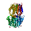
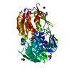
| Title | CP4 EPSPS liganded with (R)-phosphonate tetrahedral reaction intermediate analog | ||||||
 Components Components | 3-phosphoshikimate 1-carboxyvinyltransferase | ||||||
 Keywords Keywords | TRANSFERASE / inside-out alpha/beta barrel | ||||||
| Function / homology |  Function and homology information Function and homology information3-phosphoshikimate 1-carboxyvinyltransferase / 3-phosphoshikimate 1-carboxyvinyltransferase activity / chorismate biosynthetic process / response to herbicide / aromatic amino acid biosynthetic process / amino acid biosynthetic process / cytoplasm Similarity search - Function | ||||||
| Biological species |  Agrobacterium sp. (bacteria) Agrobacterium sp. (bacteria) | ||||||
| Method |  X-RAY DIFFRACTION / MOLECULAR REPLACEMENT / Resolution: 1.6 Å X-RAY DIFFRACTION / MOLECULAR REPLACEMENT / Resolution: 1.6 Å | ||||||
 Authors Authors | Healy-Fried, M.L. / Funke, T. / Han, H. / Schonbrunn, E. | ||||||
 Citation Citation | Journal: Biochemistry / Year: 2007 Title: Differential inhibition of class I and class II 5-enolpyruvylshikimate-3-phosphate synthases by tetrahedral reaction intermediate analogues. Authors: Funke, T. / Healy-Fried, M.L. / Han, H. / Alberg, D.G. / Bartlett, P.A. / Schonbrunn, E. | ||||||
| History |
|
- Structure visualization
| Structure viewer | Molecule: MolmilJmol/JSmol |
|---|
- Downloads & links
-Download
| PDBx/mmCIF format | 2pqc.cif.gz | 106.9 KB | Display | PDBx/mmCIF format |
|---|---|---|---|---|
| PDB format | pdb2pqc.ent.gz | 79.6 KB | Display | PDB format |
| PDBx/mmJSON format | 2pqc.json.gz | Tree view | PDBx/mmJSON format | |
| Others |  Other downloads Other downloads |
-Validation report
| Arichive directory | https://data.pdbj.org/pub/pdb/validation_reports/pq/2pqcftp://data.pdbj.org/pub/pdb/validation_reports/pq/2pqc | HTTPS FTP |
|---|
-Related structure data
| Related structure data |  2pq9C  2pqbC  2pqdC  2ggaS S: Starting model for refinement C: citing same article ( |
|---|---|
| Similar structure data |


-Links
 PDBj
PDBj
- Assembly
| Deposited unit | 
| ||||||||
|---|---|---|---|---|---|---|---|---|---|
| 1 |
| ||||||||
| Unit cell |
|
-Components
| #1: Protein | Mass: 46665.266 Da / Num. of mol.: 1 Source method: isolated from a genetically manipulated source Source: (gene. exp.) Agrobacterium sp. (bacteria) / Strain: CP4 / Gene: aroA / Plasmid: pET24d / Species (production host): Escherichia coli / Production host: References: UniProt: Q9R4E4, 3-phosphoshikimate 1-carboxyvinyltransferase |
|---|---|
| #2: Chemical | ChemComp-RC1 / [  Mass: 406.174 Da / Num. of mol.: 1 / Source method: obtained synthetically / Formula: C10H16O13P2 Mass: 406.174 Da / Num. of mol.: 1 / Source method: obtained synthetically / Formula: C10H16O13P2 |
| #3: Water | ChemComp-HOH /  Mass: 18.015 Da / Num. of mol.: 506 / Source method: isolated from a natural source / Formula: H2O Mass: 18.015 Da / Num. of mol.: 506 / Source method: isolated from a natural source / Formula: H2O |
-Experimental details
-Experiment
| Experiment | Method: X-RAY DIFFRACTION / Number of used crystals: 1 |
|---|
- Sample preparation
| Crystal | Density Matthews: 2.25 Å3/Da / Density % sol: 45.38 % |
|---|---|
| Crystal grow | Temperature: 290 K / Method: vapor diffusion, hanging drop / pH: 7.5 Details: (NH4)2SO4, KCl, PEG 400, HEPES-Na, pH 7.5, VAPOR DIFFUSION, HANGING DROP, temperature 290K |
-Data collection
| Diffraction | Mean temperature: 100 K |
|---|---|
| Diffraction source | Source: ROTATING ANODE / Type: RIGAKU RU300 / Wavelength: 1.5418 Å |
| Detector | Type: RIGAKU RAXIS IV / Detector: IMAGE PLATE / Date: Feb 14, 2007 / Details: mirrors |
| Radiation | Monochromator: mirrors / Protocol: SINGLE WAVELENGTH / Monochromatic (M) / Laue (L): M / Scattering type: x-ray |
| Radiation wavelength | Wavelength: 1.5418 Å / Relative weight: 1 |
| Reflection | Resolution: 1.6→15 Å / Num. all: 54614 / Num. obs: 54614 / % possible obs: 98.9 % / Observed criterion σ(F): 0 / Observed criterion σ(I): -3 / Redundancy: 3.4 % / Rmerge(I) obs: 0.069 / Net I/σ(I): 14.7 |
| Reflection shell | Resolution: 1.6→1.65 Å / Redundancy: 3 % / Rmerge(I) obs: 0.125 / Mean I/σ(I) obs: 7.2 / Num. unique all: 4686 / % possible all: 97.3 |
- Processing
| Software |
| ||||||||||||||||||||||||||||
|---|---|---|---|---|---|---|---|---|---|---|---|---|---|---|---|---|---|---|---|---|---|---|---|---|---|---|---|---|---|
| Refinement | Method to determine structure: MOLECULAR REPLACEMENT Starting model: pdb entry 2GGA Resolution: 1.6→15 Å / Cross valid method: THROUGHOUT / σ(F): 0 / σ(I): -3 / Stereochemistry target values: Engh & Huber
| ||||||||||||||||||||||||||||
| Solvent computation | Bsol: 69.482 Å2 | ||||||||||||||||||||||||||||
| Displacement parameters | Biso mean: 9.167 Å2
| ||||||||||||||||||||||||||||
| Refinement step | Cycle: LAST / Resolution: 1.6→15 Å
| ||||||||||||||||||||||||||||
| Refine LS restraints |
| ||||||||||||||||||||||||||||
| Xplor file |
|
