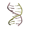
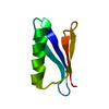
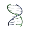
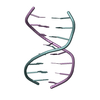
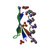
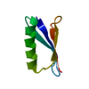
ImmunoglobulinG-bindingproteinG / IgG-binding protein G
Mass: 5941.474 Da / Num. of mol.: 1 / Fragment: First Immunoglobin binding domain (GB1) Source method: isolated from a genetically manipulated source Details: deposited only main chain atoms / Source: (gene. exp.) Streptococcus sp. 'group G' (bacteria) / Strain: group G / Gene: spg / Production host: Escherichia coli (E. coli) / References: UniProt: P19909, UniProt: P06654*PLUS
-
Experimental details
-
Experiment
Experiment
Method: SOLUTION NMR
NMR experiment
Conditions-ID
Experiment-ID
Solution-ID
Type
1
1
1
HCN-HSQC
2
2
2
water flip-back 3D 15N-edited 1H-1H COSYHMQC
-
Sample preparation
Details
Contents: perdeuterated (triple labelled 15N, 13C, 2D) Solvent system: Partially aligned in bacteriophage and lyotrophic alcohol medium.
Sample conditions
Ionic strength: 50mM salt / pH: 5.6 / Pressure: ambient / Temperature: 300 K
-
NMR measurement
NMR spectrometer
Type: Bruker AVANCE / Manufacturer: Bruker / Model: AVANCE / Field strength: 800 MHz
-
Processing
NMR software
Name
Version
Developer
Classification
Dynamic Meccano
1
bouvigniesetal
structuresolution
Dynamic Meccano
1
bouvigniesetal
refinement
Refinement
Method: dynamic meccano / Software ordinal: 1 Details: Ultrahigh-resolution backbone structure of perdeuterated protein GB1 using residual dipolar couplings from two alignment media. Angew Chem Int Ed Engl. 2006 Dec 11;45(48):8166-9.
NMR ensemble
Conformer selection criteria: all calculated structures submitted Conformers calculated total number: 1 / Conformers submitted total number: 1
+
About Yorodumi
-
News
-
Feb 9, 2022. New format data for meta-information of EMDB entries
New format data for meta-information of EMDB entries
Version 3 of the EMDB header file is now the official format.
The previous official version 1.9 will be removed from the archive.
In the structure databanks used in Yorodumi, some data are registered as the other names, "COVID-19 virus" and "2019-nCoV". Here are the details of the virus and the list of structure data.
Jan 31, 2019. EMDB accession codes are about to change! (news from PDBe EMDB page)
EMDB accession codes are about to change! (news from PDBe EMDB page)
The allocation of 4 digits for EMDB accession codes will soon come to an end. Whilst these codes will remain in use, new EMDB accession codes will include an additional digit and will expand incrementally as the available range of codes is exhausted. The current 4-digit format prefixed with “EMD-” (i.e. EMD-XXXX) will advance to a 5-digit format (i.e. EMD-XXXXX), and so on. It is currently estimated that the 4-digit codes will be depleted around Spring 2019, at which point the 5-digit format will come into force.
The EM Navigator/Yorodumi systems omit the EMD- prefix.
Related info.:Q: What is EMD? / ID/Accession-code notation in Yorodumi/EM Navigator
Yorodumi is a browser for structure data from EMDB, PDB, SASBDB, etc.
This page is also the successor to EM Navigator detail page, and also detail information page/front-end page for Omokage search.
The word "yorodu" (or yorozu) is an old Japanese word meaning "ten thousand". "mi" (miru) is to see.
Related info.:EMDB / PDB / SASBDB / Comparison of 3 databanks / Yorodumi Search / Aug 31, 2016. New EM Navigator & Yorodumi / Yorodumi Papers / Jmol/JSmol / Function and homology information / Changes in new EM Navigator and Yorodumi
 Movie
Movie Controller

 Yorodumi
 Open data
 Basic information
 Components
 Keywords
 Function and homology information
 Streptococcus sp. 'group G' (bacteria)
 Authors
 Citation
 Structure visualization
 Downloads & links
 Other downloads
 PDBj
PDBj

 Assembly
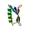
 HSQC
HSQC Sample preparation
 Processing