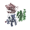
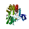
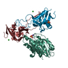
BIOMOLECULE: 1 THIS ENTRY CONTAINS THE CRYSTALLOGRAPHIC ASYMMETRIC UNIT WHICH CONSISTS OF 2 ... BIOMOLECULE: 1 THIS ENTRY CONTAINS THE CRYSTALLOGRAPHIC ASYMMETRIC UNIT WHICH CONSISTS OF 2 CHAIN(S). SEE REMARK 350 FOR INFORMATION ON GENERATING THE BIOLOGICAL MOLECULE(S). SIZE EXCLUSION CHROMATOGRAPHY SUPPORTS THE ASSIGNMENT OF A DIMER AS A SIGNIFICANT OLIGOMERIZATION STATE.
Remark 999
SEQUENCE THE CONSTRUCT WAS EXPRESSED WITH A PURIFICATION TAG MGSDKIHHHHHHENLYFQG. THE TAG WAS ... SEQUENCE THE CONSTRUCT WAS EXPRESSED WITH A PURIFICATION TAG MGSDKIHHHHHHENLYFQG. THE TAG WAS REMOVED WITH TEV PROTEASE LEAVING ONLY A GLYCINE (0) FOLLOWED BY THE TARGET SEQUENCE.
Resolution: 1.81→29.386 Å / Num. obs: 37341 / % possible obs: 82.4 % / Biso Wilson estimate: 29.733 Å2 / Rmerge(I) obs: 0.056 / Net I/σ(I): 7.74
Reflection shell
Diffraction-ID: 1
Resolution (Å)
Highest resolution (Å)
Rmerge(I) obs
Mean I/σ(I) obs
Num. measured obs
Num. unique all
% possible all
1.81-1.87
0.35
1.6
1072
3378
45.9
1.87-1.95
0.325
2
2132
4788
56.6
1.95-2.04
0.27
2.6
3968
6373
79.7
2.04-2.15
0.227
3.1
4646
6997
86.3
2.15-2.28
0.174
4
4457
6731
88.3
2.28-2.46
0.135
5
4877
7318
91.3
2.46-2.7
0.105
6.2
4682
7079
92.2
2.7-3.09
0.065
9.3
4929
7432
93.5
3.09
0.034
15.4
4910
7465
94.3
-
Phasing
Phasing
Method: MAD
-
Processing
Software
Name
Version
Classification
NB
MolProbity
3beta29
modelbuilding
SHELX
phasing
REFMAC
5.2.0019
refinement
XSCALE
datascaling
PDB_EXTRACT
2
dataextraction
MAR345
CCD
datacollection
XDS
datareduction
SHELXD
phasing
SHARP
phasing
Refinement
Method to determine structure: MAD / Resolution: 1.81→29.386 Å / Cor.coef. Fo:Fc: 0.961 / Cor.coef. Fo:Fc free: 0.937 / SU B: 7.243 / SU ML: 0.11 / TLS residual ADP flag: LIKELY RESIDUAL / Cross valid method: THROUGHOUT / σ(F): 0 / ESU R: 0.147 / ESU R Free: 0.142 Stereochemistry target values: MAXIMUM LIKELIHOOD WITH PHASES Details: 1. HYDROGENS HAVE BEEN ADDED IN RIDING POSITIONS. 2. ATOM RECORD CONTAINS RESIDUAL B FACTORS ONLY. 3. A MET-INHIBITION PROTOCOL WAS USED FOR SELENOMETHIONINE INCORPORATION DURING PROTEIN ...Details: 1. HYDROGENS HAVE BEEN ADDED IN RIDING POSITIONS. 2. ATOM RECORD CONTAINS RESIDUAL B FACTORS ONLY. 3. A MET-INHIBITION PROTOCOL WAS USED FOR SELENOMETHIONINE INCORPORATION DURING PROTEIN EXPRESSION. THE OCCUPANCY OF THE SE ATOMS IN THE MSE RESIDUES WAS REDUCED TO 0.75 TO ACCOUNT FOR THE REDUCED SCATTERING POWER DUE TO PARTIAL S-MET INCORPORATION. 4. THE SIDE CHAIN OF THE RESIDUES 46-52, 208-222 IN THE A SUBUNIT AND 60-67, 207-216 IN THE B SUBUNIT WERE DISORDERED AND WERE NOT MODELED. 5. RAMACHANDRAN OUTLIER OF RESIDUE ARG 51 IN SUBUNIT A IS LOCATED IN POOR DENSITY. 6. MAGNESIUM (MG) AND CL IONS FROM THE CRYSTALLIZATION BUFFER WERE MODELED INTO THE STRUCTURE. 7. PEG8000 FRAGMENTS (PEG) FROM CRYSTALLIZATION SOLUTION ARE MODELLED.
Rfactor
Num. reflection
% reflection
Selection details
Rfree
0.229
1871
5 %
RANDOM
Rwork
0.178
-
-
-
all
0.181
-
-
-
obs
0.181
37314
93.3 %
-
Solvent computation
Ion probe radii: 0.8 Å / Shrinkage radii: 0.8 Å / VDW probe radii: 1.2 Å / Solvent model: MASK
Displacement parameters
Biso mean: 22.525 Å2
Baniso -1
Baniso -2
Baniso -3
1-
-0.52 Å2
0 Å2
-0.14 Å2
2-
-
0.91 Å2
0 Å2
3-
-
-
-0.44 Å2
Refinement step
Cycle: LAST / Resolution: 1.81→29.386 Å
Protein
Nucleic acid
Ligand
Solvent
Total
Num. atoms
3524
0
12
255
3791
Refine LS restraints
Refine-ID
Type
Dev ideal
Dev ideal target
Number
X-RAY DIFFRACTION
r_bond_refined_d
0.017
0.022
3654
X-RAY DIFFRACTION
r_bond_other_d
0.002
0.02
2438
X-RAY DIFFRACTION
r_angle_refined_deg
1.596
1.965
4967
X-RAY DIFFRACTION
r_angle_other_deg
0.994
3
5936
X-RAY DIFFRACTION
r_dihedral_angle_1_deg
6.484
5
467
X-RAY DIFFRACTION
r_dihedral_angle_2_deg
35.071
24.26
169
X-RAY DIFFRACTION
r_dihedral_angle_3_deg
15.045
15
589
X-RAY DIFFRACTION
r_dihedral_angle_4_deg
16.283
15
27
X-RAY DIFFRACTION
r_chiral_restr
0.094
0.2
557
X-RAY DIFFRACTION
r_gen_planes_refined
0.006
0.02
4154
X-RAY DIFFRACTION
r_gen_planes_other
0.001
0.02
731
X-RAY DIFFRACTION
r_nbd_refined
0.216
0.2
815
X-RAY DIFFRACTION
r_nbd_other
0.202
0.2
2560
X-RAY DIFFRACTION
r_nbtor_refined
0.182
0.2
1805
X-RAY DIFFRACTION
r_nbtor_other
0.087
0.2
1893
X-RAY DIFFRACTION
r_xyhbond_nbd_refined
0.176
0.2
226
X-RAY DIFFRACTION
r_metal_ion_refined
0.029
0.2
2
X-RAY DIFFRACTION
r_symmetry_vdw_refined
0.278
0.2
11
X-RAY DIFFRACTION
r_symmetry_vdw_other
0.289
0.2
65
X-RAY DIFFRACTION
r_symmetry_hbond_refined
0.222
0.2
15
X-RAY DIFFRACTION
r_mcbond_it
2.234
3
2376
X-RAY DIFFRACTION
r_mcbond_other
0.628
3
948
X-RAY DIFFRACTION
r_mcangle_it
3.287
5
3716
X-RAY DIFFRACTION
r_scbond_it
5.44
8
1449
X-RAY DIFFRACTION
r_scangle_it
8.141
11
1251
Refine LS restraints NCS
Dom-ID: 1 / Auth asym-ID: A / Refine-ID: X-RAY DIFFRACTION
Ens-ID
Number
Type
Rms dev position (Å)
Weight position
1
460
LOOSEPOSITIONAL
0.31
5
1
460
LOOSETHERMAL
3.33
10
2
1750
LOOSEPOSITIONAL
0.67
5
2
1750
LOOSETHERMAL
2.82
10
3
345
LOOSEPOSITIONAL
0.52
5
3
345
LOOSETHERMAL
4.05
10
LS refinement shell
Resolution: 1.81→1.857 Å / Total num. of bins used: 20
Rfactor
Num. reflection
% reflection
Rfree
0.374
109
-
Rwork
0.263
1824
-
obs
-
1933
66.04 %
Refinement TLS params.
Method: refined / Refine-ID: X-RAY DIFFRACTION
ID
L11 (°2)
L12 (°2)
L13 (°2)
L22 (°2)
L23 (°2)
L33 (°2)
S11 (Å °)
S12 (Å °)
S13 (Å °)
S21 (Å °)
S22 (Å °)
S23 (Å °)
S31 (Å °)
S32 (Å °)
S33 (Å °)
T11 (Å2)
T12 (Å2)
T13 (Å2)
T22 (Å2)
T23 (Å2)
T33 (Å2)
Origin x (Å)
Origin y (Å)
Origin z (Å)
1
0.6842
-0.1613
0.3156
0.6327
-0.7433
1.5371
0.0228
0.0255
-0.0627
-0.0825
-0.0437
0.0169
0.1165
0.0477
0.0209
-0.0584
-0.0001
0.0046
-0.0383
-0.0186
-0.034
-4.2073
24.4004
54.1637
2
0.6432
0.0637
-0.0096
1.3401
0.4354
0.6637
0.0324
-0.1023
-0.0184
0.0337
-0.0666
-0.0185
-0.0017
-0.0187
0.0342
-0.0616
-0.0012
-0.006
-0.0224
0.0236
-0.0666
-3.0849
31.261
80.8451
Refinement TLS group
Refine-ID: X-RAY DIFFRACTION / Selection: ALL
ID
Refine TLS-ID
Auth asym-ID
Label asym-ID
Auth seq-ID
Label seq-ID
1
1
A
A
0 - 250
1 - 251
2
2
B
B
9 - 249
10 - 250
+
About Yorodumi
-
News
-
Feb 9, 2022. New format data for meta-information of EMDB entries
New format data for meta-information of EMDB entries
Version 3 of the EMDB header file is now the official format.
The previous official version 1.9 will be removed from the archive.
In the structure databanks used in Yorodumi, some data are registered as the other names, "COVID-19 virus" and "2019-nCoV". Here are the details of the virus and the list of structure data.
Jan 31, 2019. EMDB accession codes are about to change! (news from PDBe EMDB page)
EMDB accession codes are about to change! (news from PDBe EMDB page)
The allocation of 4 digits for EMDB accession codes will soon come to an end. Whilst these codes will remain in use, new EMDB accession codes will include an additional digit and will expand incrementally as the available range of codes is exhausted. The current 4-digit format prefixed with “EMD-” (i.e. EMD-XXXX) will advance to a 5-digit format (i.e. EMD-XXXXX), and so on. It is currently estimated that the 4-digit codes will be depleted around Spring 2019, at which point the 5-digit format will come into force.
The EM Navigator/Yorodumi systems omit the EMD- prefix.
Related info.:Q: What is EMD? / ID/Accession-code notation in Yorodumi/EM Navigator
Yorodumi is a browser for structure data from EMDB, PDB, SASBDB, etc.
This page is also the successor to EM Navigator detail page, and also detail information page/front-end page for Omokage search.
The word "yorodu" (or yorozu) is an old Japanese word meaning "ten thousand". "mi" (miru) is to see.
Related info.:EMDB / PDB / SASBDB / Comparison of 3 databanks / Yorodumi Search / Aug 31, 2016. New EM Navigator & Yorodumi / Yorodumi Papers / Jmol/JSmol / Function and homology information / Changes in new EM Navigator and Yorodumi
 Movie
Movie Controller

 Yorodumi
 Open data
 Basic information
 Components
 Keywords
 Function and homology information
 Xanthomonas campestris pv. campestris (bacteria)
 X-RAY DIFFRACTION /
  Authors
 Citation
 Structure visualization
 Downloads & links
 Other downloads
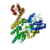


 PDBj
PDBj

 Assembly

 Mass: 24.305 Da / Num. of mol.: 2 / Source method: obtained synthetically / Formula: Mg
 Mass: 35.453 Da / Num. of mol.: 3 / Source method: obtained synthetically / Formula: Cl
 Mass: 106.120 Da / Num. of mol.: 1 / Source method: obtained synthetically / Formula: C4H10O3
 Mass: 18.015 Da / Num. of mol.: 255 / Source method: isolated from a natural source / Formula: H2O
 Sample preparation
 / Beamline: BL9-2 / Wavelength: 0.91162, 0.97936
 Processing