 Movie
Movie Controller

[English] 日本語
 Yorodumi
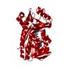
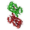
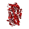
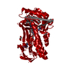
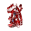
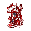
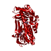
- PDB-2phh: THE COENZYME ANALOGUE ADENOSINE 5-DIPHOSPHORIBOSE DISPLACES FAD I... -
+ Open data

- Basic information
| Entry | Database: PDB / ID: 2phh | ||||||
|---|---|---|---|---|---|---|---|
| Title | THE COENZYME ANALOGUE ADENOSINE 5-DIPHOSPHORIBOSE DISPLACES FAD IN THE ACTIVE SITE OF P-HYDROXYBENZOATE HYDROXYLASE. AN X-RAY CRYSTALLOGRAPHIC INVESTIGATION | ||||||
 Components Components | P-HYDROXYBENZOATE HYDROXYLASE | ||||||
 Keywords Keywords | OXIDOREDUCTASE | ||||||
| Function / homology |  Function and homology information Function and homology information4-hydroxybenzoate 3-monooxygenase (NADPH) activity / 4-hydroxybenzoate 3-monooxygenase / 4-hydroxybenzoate 3-monooxygenase activity / : / FAD binding / flavin adenine dinucleotide binding Similarity search - Function | ||||||
| Biological species |  Pseudomonas fluorescens (bacteria) Pseudomonas fluorescens (bacteria) | ||||||
| Method |  X-RAY DIFFRACTION / Resolution: 2.7 Å X-RAY DIFFRACTION / Resolution: 2.7 Å | ||||||
 Authors Authors | Van Derlaan, J.M. / Drenth, J. / Hol, W.G.J. | ||||||
 Citation Citation | Journal: Biochemistry / Year: 1989 Title: The coenzyme analogue adenosine 5-diphosphoribose displaces FAD in the active site of p-hydroxybenzoate hydroxylase. An x-ray crystallographic investigation. Authors: van der Laan, J.M. / Schreuder, H.A. / Swarte, M.B. / Wierenga, R.K. / Kalk, K.H. / Hol, W.G. / Drenth, J. #1: Journal: Eur.J.Biochem. / Year: 1989Title: The Influence of Purification and Protein Heterogeneity on the Crystallization of P-Hydroxybenzoate Hydroxylase Authors: Van Derlaan, J.M. / Swarte, M.B.A. / Groendijk, H. / Hol, W.G.J. / Drenth, J. #2: Journal: J.Mol.Biol. / Year: 1989Title: Crystal Structure of the P-Hydroxylase-Substrate Complex Refined at 1.9 Angstroms Resolution Authors: Schreuder, H.A. / Prick, P.A.J. / Wierenga, R.K. / Vriend, G. / Wilson, K.S. / Hol, W.G.J. / Drenth, J. #3: Journal: J.Mol.Biol. / Year: 1988Title: Crystal Structure of P-Hydroxybenzoate Hydroxylas Complexed with its Reaction Product 3,4-Dihydroxybenzoate Authors: Schreuder, H.A. / Van Derlaan, J.M. / Hol, W.G.J. / Drenth, J. #4: Journal: Eur.J.Biochem. / Year: 1983Title: P-Hydroxybenzoate Hydroxylase from Pseudomonas Fluorescens. 1. Completion of the Elucidation of the Primary Structure Authors: Hofsteenge, J. / Weijer, W.J. / Jekel, P.A. / Beintema, J.J. #5: Journal: Eur.J.Biochem. / Year: 1983Title: P-Hydroxybenzoate Hydroxylase from Pseudomonas Fluorescens. 2. Fitting of the Amino-Acid Sequence to the Tertiary Structure Authors: Weijer, W.J. / Hofsteenge, J. / Beintema, J.J. / Wierenga, R.K. / Drenth, J. #6: Journal: J.Mol.Biol. / Year: 1983Title: Comparison of the Three-Dimensional Protein and Nucleotide Structure of the Fad-Binding Domain of P-Hydroxybenzoate Hydroxylase with the Fad-as Well as Nadph-Binding Domains of Glutathione Reductase Authors: Wierenga, R.K. / Drenth, J. / Schulz, G.E. #7: Journal: Eur.J.Biochem. / Year: 1980Title: Primary and Tertiary Structure Studies of P-Hydroxybenzoate Hydroxylase from Pseudomonas Fluorescens. Isolation and Alignment of the Cnbr Peptides. Interactions of the Protein with Flavin Adenine Dinucleotide Authors: Hofsteenge, J. / Vereijken, J.M. / Weijer, W.J. / Beintema, J.J. / Wierenga, R.K. / Drenth, J. #8: Journal: Eur.J.Biochem. / Year: 1980Title: The Amino-Acid Sequence of the Three Smallest Cnbr Peptides from P-Hydroxybenzoate Hydroxylase from Pseudomonas Florescens Authors: Vereijken, J.M. / Hofsteenge, J. / Bak, H.J. / Beintema, J.J. #9: Journal: J.Mol.Biol. / Year: 1979Title: Crystal Structure of P-Hydroxybenzoate Hydroxylase Authors: Wierenga, R.K. / Dejong, R.J. / Kalk, K.H. / Hol, W.G.J. / Drenth, J. #10: Journal: J.Biol.Chem. / Year: 1975Title: Crystallization and Preliminary X-Ray Investigation of P-Hydrobenzoate Hydroxylase from Pseudomonas Fluorescens Authors: Drenth, J. / Hol, W.G.J. / Wierenga, R.K. | ||||||
| History |
|
- Structure visualization
| Structure viewer | Molecule: MolmilJmol/JSmol |
|---|
- Downloads & links
-Download
| PDBx/mmCIF format | 2phh.cif.gz | 92.8 KB | Display | PDBx/mmCIF format |
|---|---|---|---|---|
| PDB format | pdb2phh.ent.gz | 70.4 KB | Display | PDB format |
| PDBx/mmJSON format | 2phh.json.gz | Tree view | PDBx/mmJSON format | |
| Others |  Other downloads Other downloads |
-Validation report
| Arichive directory | https://data.pdbj.org/pub/pdb/validation_reports/ph/2phhftp://data.pdbj.org/pub/pdb/validation_reports/ph/2phh | HTTPS FTP |
|---|
-Related structure data
| Similar structure data |
|---|
-Links
 PDBj
PDBj
- Assembly
| Deposited unit | 
| ||||||||
|---|---|---|---|---|---|---|---|---|---|
| 1 | 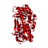
| ||||||||
| Unit cell |
| ||||||||
| Atom site foot note | 1: RESIDUE 275 IS A CIS-PROLINE. |
-Components
| #1: Protein | Mass: 44380.574 Da / Num. of mol.: 1 Source method: isolated from a genetically manipulated source Source: (gene. exp.) Pseudomonas fluorescens (bacteria)References: UniProt: P00438, 4-hydroxybenzoate 3-monooxygenase |
|---|---|
| #2: Chemical | ChemComp-APR /   Mass: 559.316 Da / Num. of mol.: 1 / Source method: obtained synthetically / Formula: C15H23N5O14P2 Mass: 559.316 Da / Num. of mol.: 1 / Source method: obtained synthetically / Formula: C15H23N5O14P2 |
| #3: Chemical | ChemComp-PHB / 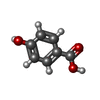  Mass: 138.121 Da / Num. of mol.: 1 / Source method: obtained synthetically / Formula: C7H6O3 Mass: 138.121 Da / Num. of mol.: 1 / Source method: obtained synthetically / Formula: C7H6O3 |
| #4: Water | ChemComp-HOH /  Mass: 18.015 Da / Num. of mol.: 57 / Source method: isolated from a natural source / Formula: H2O Mass: 18.015 Da / Num. of mol.: 57 / Source method: isolated from a natural source / Formula: H2O |
-Experimental details
-Experiment
| Experiment | Method: X-RAY DIFFRACTION |
|---|
- Sample preparation
| Crystal | Density Matthews: 2.64 Å3/Da / Density % sol: 53.34 % | ||||||||||||||||||||||||||||||||||||||||||||||||||||||||||||||||||||||||||||||||||||||||||
|---|---|---|---|---|---|---|---|---|---|---|---|---|---|---|---|---|---|---|---|---|---|---|---|---|---|---|---|---|---|---|---|---|---|---|---|---|---|---|---|---|---|---|---|---|---|---|---|---|---|---|---|---|---|---|---|---|---|---|---|---|---|---|---|---|---|---|---|---|---|---|---|---|---|---|---|---|---|---|---|---|---|---|---|---|---|---|---|---|---|---|---|
| Crystal grow | *PLUS pH: 7.5 / Method: unknown | ||||||||||||||||||||||||||||||||||||||||||||||||||||||||||||||||||||||||||||||||||||||||||
| Components of the solutions | *PLUS
|
-Data collection
| Reflection | *PLUS Highest resolution: 2.7 Å / Num. obs: 9857 / % possible obs: 70 % / Num. measured all: 19604 / Rmerge(I) obs: 0.066 |
|---|
- Processing
| Software | Name: PROLSQ / Classification: refinement | ||||||||||||||||||||||||||||||||||||||||||||||||||||||||||||||||||||||||||||||||||||
|---|---|---|---|---|---|---|---|---|---|---|---|---|---|---|---|---|---|---|---|---|---|---|---|---|---|---|---|---|---|---|---|---|---|---|---|---|---|---|---|---|---|---|---|---|---|---|---|---|---|---|---|---|---|---|---|---|---|---|---|---|---|---|---|---|---|---|---|---|---|---|---|---|---|---|---|---|---|---|---|---|---|---|---|---|---|
| Refinement | Rfactor obs: 0.168 / Highest resolution: 2.7 Å | ||||||||||||||||||||||||||||||||||||||||||||||||||||||||||||||||||||||||||||||||||||
| Refinement step | Cycle: LAST / Highest resolution: 2.7 Å
| ||||||||||||||||||||||||||||||||||||||||||||||||||||||||||||||||||||||||||||||||||||
| Refine LS restraints |
| ||||||||||||||||||||||||||||||||||||||||||||||||||||||||||||||||||||||||||||||||||||
| Refinement | *PLUS Highest resolution: 2.7 Å / Lowest resolution: 6 Å / Rfactor obs: 0.168 / Num. reflection obs: 8836 | ||||||||||||||||||||||||||||||||||||||||||||||||||||||||||||||||||||||||||||||||||||
| Solvent computation | *PLUS | ||||||||||||||||||||||||||||||||||||||||||||||||||||||||||||||||||||||||||||||||||||
| Displacement parameters | *PLUS |
