 Movie
Movie Controller

[English] 日本語
 Yorodumi
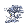
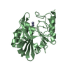
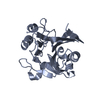
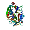
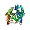
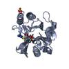
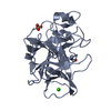
- PDB-2pfw: CRYSTAL STRUCTURE OF A RMLC-LIKE CUPIN (SFRI_3105) FROM SHEWANELL... -
+ Open data

- Basic information
| Entry | Database: PDB / ID: 2pfw | ||||||
|---|---|---|---|---|---|---|---|
| Title | CRYSTAL STRUCTURE OF A RMLC-LIKE CUPIN (SFRI_3105) FROM SHEWANELLA FRIGIDIMARINA NCIMB 400 AT 1.90 A RESOLUTION | ||||||
 Components Components | Cupin 2, conserved barrel domain protein | ||||||
 Keywords Keywords | UNKNOWN FUNCTION / CUPIN DOMAIN / CUPIN 2 CONSERVED BARREL DOMAIN PROTEIN / STRUCTURAL GENOMICS / JOINT CENTER FOR STRUCTURAL GENOMICS / JCSG / PROTEIN STRUCTURE INITIATIVE / PSI-2 | ||||||
| Function / homology |  Function and homology information Function and homology informationPectin degradation protein KdgF / : / Cupin 2, conserved barrel / Cupin domain / RmlC-like cupin domain superfamily / Jelly Rolls / RmlC-like jelly roll fold / Jelly Rolls / Sandwich / Mainly Beta Similarity search - Domain/homology | ||||||
| Biological species |  Shewanella frigidimarina (bacteria) Shewanella frigidimarina (bacteria) | ||||||
| Method |  X-RAY DIFFRACTION / SYNCHROTRON / MAD / Resolution: 1.9 Å X-RAY DIFFRACTION / SYNCHROTRON / MAD / Resolution: 1.9 Å | ||||||
 Authors Authors | Joint Center for Structural Genomics (JCSG) | ||||||
 Citation Citation | Journal: To be published Title: Crystal structure of Cupin 2 conserved barrel domain protein (YP_751781.1) from Shewanella frigidimarina NCIMB 400 at 1.90 A resolution Authors: Joint Center for Structural Genomics (JCSG) | ||||||
| History |
| ||||||
| Remark 300 | BIOMOLECULE: 1 THIS ENTRY CONTAINS THE CRYSTALLOGRAPHIC ASYMMETRIC UNIT WHICH CONSISTS OF 1 CHAINS. ... BIOMOLECULE: 1 THIS ENTRY CONTAINS THE CRYSTALLOGRAPHIC ASYMMETRIC UNIT WHICH CONSISTS OF 1 CHAINS. SEE REMARK 350 FOR INFORMATION ON GENERATING THE BIOLOGICAL MOLECULE(S). SIZE EXCLUSION CHROMATOGRAPHY WITH STATIC LIGHT SCATTERING SUPPORTS THE ASSIGNMENT OF A DIMER AS A BIOLOGICALLY SIGNIFICANT OLIGOMERIZATION STATE. | ||||||
| Remark 999 | SEQUENCE THE CONSTRUCT WAS EXPRESSED WITH A PURIFICATION TAG MGSDKIHHHHHHENLYFQG. THE TAG WAS ... SEQUENCE THE CONSTRUCT WAS EXPRESSED WITH A PURIFICATION TAG MGSDKIHHHHHHENLYFQG. THE TAG WAS REMOVED WITH TEV PROTEASE LEAVING ONLY A GLYCINE (0) FOLLOWED BY THE TARGET SEQUENCE. |
- Structure visualization
| Structure viewer | Molecule: MolmilJmol/JSmol |
|---|
- Downloads & links
-Download
| PDBx/mmCIF format | 2pfw.cif.gz | 61.2 KB | Display | PDBx/mmCIF format |
|---|---|---|---|---|
| PDB format | pdb2pfw.ent.gz | 44 KB | Display | PDB format |
| PDBx/mmJSON format | 2pfw.json.gz | Tree view | PDBx/mmJSON format | |
| Others |  Other downloads Other downloads |
-Validation report
| Arichive directory | https://data.pdbj.org/pub/pdb/validation_reports/pf/2pfwftp://data.pdbj.org/pub/pdb/validation_reports/pf/2pfw | HTTPS FTP |
|---|
-Related structure data
| Similar structure data | |
|---|---|
| Other databases |


-Links
 PDBj
PDBj- Assembly
| Deposited unit | 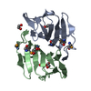
| ||||||||
|---|---|---|---|---|---|---|---|---|---|
| 1 |
| ||||||||
| Unit cell |
| ||||||||
| Details | SIZE EXCLUSION CHROMATOGRAPHY WITH STATIC LIGHT SCATTERING SUPPORTS THE ASSIGNMENT OF A DIMER AS A BIOLOGICALLY SIGNIFICANT OLIGOMERIZATION STATE. |
-Components
| #1: Protein | Mass: 12882.890 Da / Num. of mol.: 2 Source method: isolated from a genetically manipulated source Source: (gene. exp.) Shewanella frigidimarina (bacteria) / Strain: NCIMB 400 / Gene: YP_751781.1, Sfri_3105 / Plasmid: speedET / Production host: #2: Chemical | ChemComp-EDO /   Mass: 62.068 Da / Num. of mol.: 5 / Source method: obtained synthetically / Formula: C2H6O2 Mass: 62.068 Da / Num. of mol.: 5 / Source method: obtained synthetically / Formula: C2H6O2#3: Water | ChemComp-HOH / |  Mass: 18.015 Da / Num. of mol.: 111 / Source method: isolated from a natural source / Formula: H2O Mass: 18.015 Da / Num. of mol.: 111 / Source method: isolated from a natural source / Formula: H2OHas protein modification | Y | |
|---|
-Experimental details
-Experiment
| Experiment | Method: X-RAY DIFFRACTION / Number of used crystals: 1 |
|---|
- Sample preparation
| Crystal | Density Matthews: 3.04 Å3/Da / Density % sol: 59.47 % Description: THE STRUCTURE WAS SOLVED IN SPACE GROUP P41212. MAD PHASING STATISTICS AND AUTOMATED MODEL BUILDING LOOKED REASONABLE. AFTER INITIAL REFINEMENT IN P41212, THE R AND FREE-R FACTORS WERE ...Description: THE STRUCTURE WAS SOLVED IN SPACE GROUP P41212. MAD PHASING STATISTICS AND AUTOMATED MODEL BUILDING LOOKED REASONABLE. AFTER INITIAL REFINEMENT IN P41212, THE R AND FREE-R FACTORS WERE HIGHER THAN EXPECTED. THE STRUCTURE WAS THEN EXPANDED TO SPACEGROUPS P41 AND P212121. TWINNING TESTS IN BOTH SPACE GROUPS YIELDED IMPROVED STATISTICS. HOWEVER, ONLY THE P212121 TWIN REFINEMENT GAVE THE EXPECTED R/FREE-R VALUES. SUBSEQUENTLY, THE MAD PHASING WAS REPEATED IN SPACE GROUP P212121. THE REPORTED STATISTICS REFER TO THE REFINEMENT AGAINST THE TWINNED DATA. |
|---|---|
| Crystal grow | Temperature: 277 K / Method: vapor diffusion, sitting drop / pH: 7.5 Details: NANODROP, 1.4M Na3Citrate, 0.1M HEPES pH 7.5, VAPOR DIFFUSION, SITTING DROP, temperature 277K |
-Data collection
| Diffraction | Mean temperature: 100 K | |||||||||||||||||||||||||||||||||||||||||||||||||||||||||||||||||||||||||||||
|---|---|---|---|---|---|---|---|---|---|---|---|---|---|---|---|---|---|---|---|---|---|---|---|---|---|---|---|---|---|---|---|---|---|---|---|---|---|---|---|---|---|---|---|---|---|---|---|---|---|---|---|---|---|---|---|---|---|---|---|---|---|---|---|---|---|---|---|---|---|---|---|---|---|---|---|---|---|---|
| Diffraction source | Source: SYNCHROTRON / Site: SSRL  / Beamline: BL11-1 / Wavelength: 0.91162, 0.97895, 0.97871 / Beamline: BL11-1 / Wavelength: 0.91162, 0.97895, 0.97871 | |||||||||||||||||||||||||||||||||||||||||||||||||||||||||||||||||||||||||||||
| Detector | Type: MARMOSAIC 325 mm CCD / Detector: CCD / Date: Jan 18, 2007 / Details: Flat mirror (vertical focusing) | |||||||||||||||||||||||||||||||||||||||||||||||||||||||||||||||||||||||||||||
| Radiation | Monochromator: Single crystal Si(111) bent (horizontal focusing) Protocol: MAD / Monochromatic (M) / Laue (L): M / Scattering type: x-ray | |||||||||||||||||||||||||||||||||||||||||||||||||||||||||||||||||||||||||||||
| Radiation wavelength |
| |||||||||||||||||||||||||||||||||||||||||||||||||||||||||||||||||||||||||||||
| Reflection | Resolution: 1.9→29.248 Å / Num. obs: 42979 / % possible obs: 89.8 % / Biso Wilson estimate: 28.762 Å2 / Rmerge(I) obs: 0.052 / Net I/σ(I): 12.26 | |||||||||||||||||||||||||||||||||||||||||||||||||||||||||||||||||||||||||||||
| Reflection shell |
|
-Phasing
| Phasing | Method: MAD |
|---|
- Processing
| Software |
| ||||||||||||||||||||||||||||||||||||||||
|---|---|---|---|---|---|---|---|---|---|---|---|---|---|---|---|---|---|---|---|---|---|---|---|---|---|---|---|---|---|---|---|---|---|---|---|---|---|---|---|---|---|
| Refinement | Method to determine structure: MAD / Resolution: 1.9→29.248 Å / Num. parameters: 7524 / Num. restraintsaints: 19194 / Cross valid method: FREE R / σ(F): 0 / Stereochemistry target values: ENGH AND HUBER Details: 1. HYDROGENS HAVE BEEN ADDED IN THE RIDING POSITIONS. 2. RESIDUES 113-115 IN CHAIN A AND 112-115 IN CHAIN B ARE DISORDERED AND ARE NOT MODELED. 3. REFINEMENT WAS PERFORMED AGAINST INTENSITY ...Details: 1. HYDROGENS HAVE BEEN ADDED IN THE RIDING POSITIONS. 2. RESIDUES 113-115 IN CHAIN A AND 112-115 IN CHAIN B ARE DISORDERED AND ARE NOT MODELED. 3. REFINEMENT WAS PERFORMED AGAINST INTENSITY DATA. 4. UNMERGED INTENSITY DATA WAS MERGED WITH SHELXL MERG 2 OPTION (FRIEDEL PAIRS SEPARATE). 5. THE SE FORM FACTORS WERE MODIFIED WITH f' -1.8 and f" 3.4 6. THE DATA ARE PSEUDO-MEROHEDRALLY TWINNED IN SPACE GROUP P212121 WITH THE TWIN LAW -H,L,K. THE REFINED TWIN FRACTION WAS 0.47.
| ||||||||||||||||||||||||||||||||||||||||
| Solvent computation | Solvent model: MOEWS & KRETSINGER, J.MOL.BIOL.91(1973)201-228 | ||||||||||||||||||||||||||||||||||||||||
| Displacement parameters | Biso mean: 26.904 Å2 | ||||||||||||||||||||||||||||||||||||||||
| Refine analyze | Num. disordered residues: 1 / Occupancy sum hydrogen: 1580 / Occupancy sum non hydrogen: 1874 | ||||||||||||||||||||||||||||||||||||||||
| Refinement step | Cycle: LAST / Resolution: 1.9→29.248 Å
| ||||||||||||||||||||||||||||||||||||||||
| Refine LS restraints |
|
