 Movie
Movie Controller

[English] 日本語
 Yorodumi
- PDB-2pfk: THE CRYSTAL STRUCTURE OF UNLIGANDED PHOSPHOFRUCTOKINASE FROM ESCH... -
+ Open data

- Basic information
| Entry | Database: PDB / ID: 2pfk | |||||||||
|---|---|---|---|---|---|---|---|---|---|---|
| Title | THE CRYSTAL STRUCTURE OF UNLIGANDED PHOSPHOFRUCTOKINASE FROM ESCHERICHIA COLI | |||||||||
 Components Components | 6-PHOSPHOFRUCTOKINASE ISOZYME I | |||||||||
 Keywords Keywords | TRANSFERASE(PHOSPHOTRANSFERASE) | |||||||||
| Function / homology |  Function and homology information Function and homology information6-phosphofructokinase complex / 6-phosphofructokinase / ribonucleotide binding / 6-phosphofructokinase activity / fructose-6-phosphate binding / glucose catabolic process / fructose 1,6-bisphosphate metabolic process / fructose 6-phosphate metabolic process / canonical glycolysis / glycolytic process ...6-phosphofructokinase complex / 6-phosphofructokinase / ribonucleotide binding / 6-phosphofructokinase activity / fructose-6-phosphate binding / glucose catabolic process / fructose 1,6-bisphosphate metabolic process / fructose 6-phosphate metabolic process / canonical glycolysis / glycolytic process / GDP binding / protein homotetramerization / magnesium ion binding / ATP binding / identical protein binding / cytoplasm / cytosol Similarity search - Function | |||||||||
| Biological species |  | |||||||||
| Method |  X-RAY DIFFRACTION / Resolution: 2.4 Å X-RAY DIFFRACTION / Resolution: 2.4 Å | |||||||||
 Authors Authors | Rypniewski, W.R. / Evans, P.R. | |||||||||
 Citation Citation | Journal: J.Mol.Biol. / Year: 1989 Title: Crystal structure of unliganded phosphofructokinase from Escherichia coli. Authors: Rypniewski, W.R. / Evans, P.R. #1: Journal: J.Mol.Biol. / Year: 1988Title: Crystal Structure of the Complex of Phosphofructokinase from Escherichia Coli with its Reaction Products Authors: Shirakihara, Y. / Evans, P.R. #2: Journal: J.Mol.Biol. / Year: 1986Title: Crystallographic Structure of Allosterically Inhibited Phosphofructokinase at 7 Angstroms Resolution Authors: Evans, P.R. / Farrants, G.W. / Lawrence, M.C. #3: Journal: Eur.J.Biochem. / Year: 1985Title: Nucleotide Sequence and High-Level Expression of the Major Escherichia Coli Phosphofructokinase Authors: Hellinga, H.W. / Evans, P.R. #4: Journal: Philos.Trans.R.Soc.London,Ser.B / Year: 1981Title: Phosphofructokinase. Structure and Control Authors: Evans, P.R. / Farrants, G.W. / Hudson, P.J. #5: Journal: Nature / Year: 1979Title: Structure and Control of Phosphofructokinase from Bacillus Stearothermophilus Authors: Evans, P.R. / Hudson, P.J. #6: Journal: Proc.FEBS Meet. / Year: 1978Title: The Three-Dimensional Structure of Phosphofructokinase from Bacillus Stearothermophilus Authors: Evans, P.R. / Hudson, P.J. | |||||||||
| History |
| |||||||||
| Remark 285 | THE ENTRY COORDINATES ARE NOT PRESENTED IN THE STANDARD CRYSTAL FRAME. |
- Structure visualization
| Structure viewer | Molecule: MolmilJmol/JSmol |
|---|
- Downloads & links
-Download
| PDBx/mmCIF format | 2pfk.cif.gz | 239.3 KB | Display | PDBx/mmCIF format |
|---|---|---|---|---|
| PDB format | pdb2pfk.ent.gz | 193.7 KB | Display | PDB format |
| PDBx/mmJSON format | 2pfk.json.gz | Tree view | PDBx/mmJSON format | |
| Others |  Other downloads Other downloads |
-Validation report
| Arichive directory | https://data.pdbj.org/pub/pdb/validation_reports/pf/2pfkftp://data.pdbj.org/pub/pdb/validation_reports/pf/2pfk | HTTPS FTP |
|---|
-Related structure data
| Similar structure data |
|---|
-Links
 PDBj
PDBj
- Assembly
| Deposited unit | 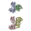
| ||||||||||||||||
|---|---|---|---|---|---|---|---|---|---|---|---|---|---|---|---|---|---|
| 1 | 
| ||||||||||||||||
| 2 | 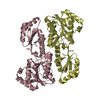
| ||||||||||||||||
| Unit cell |
| ||||||||||||||||
| Components on special symmetry positions |
| ||||||||||||||||
| Noncrystallographic symmetry (NCS) | NCS oper:
|
-Components
| #1: Protein | Mass: 34885.047 Da / Num. of mol.: 4 Source method: isolated from a genetically manipulated source Source: (gene. exp.) References: PIR: KIECFA, UniProt: P0A796*PLUS, 6-phosphofructokinase #2: Water | ChemComp-HOH / |  Mass: 18.015 Da / Num. of mol.: 347 / Source method: isolated from a natural source / Formula: H2O Mass: 18.015 Da / Num. of mol.: 347 / Source method: isolated from a natural source / Formula: H2O |
|---|
-Experimental details
-Experiment
| Experiment | Method: X-RAY DIFFRACTION |
|---|
- Sample preparation
| Crystal | Density Matthews: 2.84 Å3/Da / Density % sol: 56.7 % | ||||||||||||||||||||||||||||||
|---|---|---|---|---|---|---|---|---|---|---|---|---|---|---|---|---|---|---|---|---|---|---|---|---|---|---|---|---|---|---|---|
| Crystal grow | *PLUS Method: batch method / PH range low: 7.9 / PH range high: 7.7 | ||||||||||||||||||||||||||||||
| Components of the solutions | *PLUS
|
-Data collection
| Reflection | *PLUS Highest resolution: 2.4 Å / Num. obs: 59481 / Num. measured all: 388304 / Rmerge(I) obs: 0.057 |
|---|---|
| Reflection shell | *PLUS Highest resolution: 2.4 Å / Lowest resolution: 2.6 Å |
- Processing
| Software | Name: PROLSQ / Classification: refinement | |||||||||||||||||||||||||||||||||||||||||||||||||||||||||||||||
|---|---|---|---|---|---|---|---|---|---|---|---|---|---|---|---|---|---|---|---|---|---|---|---|---|---|---|---|---|---|---|---|---|---|---|---|---|---|---|---|---|---|---|---|---|---|---|---|---|---|---|---|---|---|---|---|---|---|---|---|---|---|---|---|---|
| Refinement | Resolution: 2.4→100 Å / Rfactor obs: 0.166 Details: THE SPACE GROUP HAS BEEN CONSIDERED AS C 21, I.E. C 2 WITH THE ORIGIN ON A 21 AXIS. THIS IS THE SAME AS P 21 PLUS THE C-CENTERING. THE COORDINATES IN THIS ENTRY ARE IN THE A*, B, C ...Details: THE SPACE GROUP HAS BEEN CONSIDERED AS C 21, I.E. C 2 WITH THE ORIGIN ON A 21 AXIS. THIS IS THE SAME AS P 21 PLUS THE C-CENTERING. THE COORDINATES IN THIS ENTRY ARE IN THE A*, B, C ORTHOGONAL COORDINATE FRAME. THE CRYSTALLOGRAPHIC SYMMETRY OPERATIONS ARE (X, Y, Z), (-X, 1/2+Y, -Z), (1/2+X, 1/2+Y, Z), AND (1/2-X, Y, -Z). THE ASYMMETRIC UNIT CONTAINS TWO HALF-TETRAMERS, I. E. THERE ARE TWO DIFFERENT SORTS OF TETRAMERS WHICH SIT ON DIFFERENT CRYSTALLOGRAPHIC DYAD AXES, AT (-1/4, 0, 0) AND (1/4, 0, 1/2). THUS THERE ARE FOUR DIFFERENT SUBUNITS IN THE UNIT CELL. THESE SUBUNITS HAVE BEEN ASSIGNED CHAIN IDENTIFIERS A, B, C, AND D. SUBUNITS A AND B ARE RELATED BY A PSEUDO-DYAD. THE TRANSFORMATION PRESENTED ON THE *MTRIX 1* RECORDS BELOW WILL YIELD COORDINATES FOR CHAIN B WHEN APPLIED TO CHAIN A AND VICE VERSA. SUBUNITS C AND D ARE RELATED BY A PSEUDO-DYAD. THE TRANSFORMATION PRESENTED ON THE *MTRIX 2* RECORDS BELOW WILL YIELD COORDINATES FOR CHAIN D WHEN APPLIED TO CHAIN C AND VICE VERSA. TO GENERATE A TETRAMER FROM SUBUNITS A AND B ONE MUST APPLY THE FOLLOWING CRYSTALLOGRAPHIC SYMMETRY OPERATION TO THE COORDINATES PRESENTED BELOW FOR CHAINS A AND B - -1.0 0.0 0.0 -77.53963 0.0 1.0 0.0 0.0 0.0 0.0 -1.0 42.68050 TO GENERATE A TETRAMER FROM SUBUNITS C AND D ONE MUST APPLY THE FOLLOWING CRYSTALLOGRAPHIC SYMMETRY OPERATION TO THE COORDINATES PRESENTED BELOW FOR CHAINS C AND D - -1.0 0.0 0.0 77.53963 0.0 1.0 0.0 0.0 0.0 0.0 -1.0 111.28951 THE TETRAMER FORMED FROM SUBUNITS A AND B IS CENTERED AT (-1/4, 0, 0) AND THE TETRAMER FORMED FROM SUBUNITS C AND D IS CENTERED AT (1/4, 20.89/66.4, 1/2). THE SUBUNITS ARE ALL SIMILAR IN CONFORMATION. THE TRANSFORMATION PRESENTED ON THE *MTRIX 3* RECORDS BELOW WILL YIELD COORDINATES FOR CHAIN C WHEN APPLIED TO CHAIN A. ALL OTHER TRANSFORMATIONS BETWEEN CHAINS CAN BE GENERATED FROM THE GIVEN TRANSFORMATIONS. | |||||||||||||||||||||||||||||||||||||||||||||||||||||||||||||||
| Refinement step | Cycle: LAST / Resolution: 2.4→100 Å
| |||||||||||||||||||||||||||||||||||||||||||||||||||||||||||||||
| Refine LS restraints |
| |||||||||||||||||||||||||||||||||||||||||||||||||||||||||||||||
| Software | *PLUS Name: PROLSQ / Classification: refinement | |||||||||||||||||||||||||||||||||||||||||||||||||||||||||||||||
| Refinement | *PLUS Rfactor obs: 0.166 | |||||||||||||||||||||||||||||||||||||||||||||||||||||||||||||||
| Solvent computation | *PLUS | |||||||||||||||||||||||||||||||||||||||||||||||||||||||||||||||
| Displacement parameters | *PLUS | |||||||||||||||||||||||||||||||||||||||||||||||||||||||||||||||
| Refine LS restraints | *PLUS
|
