 Movie
Movie Controller

[English] 日本語
 Yorodumi
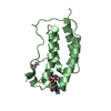
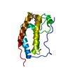
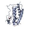
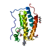
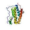
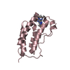
- PDB-2pf1: STRUCTURE OF BOVINE PROTHROMBIN FRAGMENT 1 REFINED AT 2.25 ANGSTR... -
+ Open data

- Basic information
| Entry | Database: PDB / ID: 2pf1 | ||||||
|---|---|---|---|---|---|---|---|
| Title | STRUCTURE OF BOVINE PROTHROMBIN FRAGMENT 1 REFINED AT 2.25 ANGSTROMS RESOLUTION | ||||||
 Components Components | PROTHROMBIN FRAGMENT 1 | ||||||
 Keywords Keywords | HYDROLASE(SERINE PROTEINASE) | ||||||
| Function / homology |  Function and homology information Function and homology informationfibrinogen binding / thrombin / positive regulation of blood coagulation / protein polymerization / acute-phase response / platelet activation / serine-type endopeptidase activity / calcium ion binding / proteolysis / : Similarity search - Function | ||||||
| Biological species |  | ||||||
| Method |  X-RAY DIFFRACTION / Resolution: 2.8 Å X-RAY DIFFRACTION / Resolution: 2.8 Å | ||||||
 Authors Authors | Seshadri, T.P. / Tulinsky, A. / Skrzypczak-Jankun, E. / Park, C.H. | ||||||
 Citation Citation | Journal: J.Mol.Biol. / Year: 1991 Title: Structure of bovine prothrombin fragment 1 refined at 2.25 A resolution. Authors: Seshadri, T.P. / Tulinsky, A. / Skrzypczak-Jankun, E. / Park, C.H. #1: Journal: J.Mol.Biol. / Year: 1988Title: Structure of Prothrombin Fragment 1 Refined at 2.8 Angstroms Resolution Authors: Tulinsky, A. / Park, C.H. / Skrzypczak-Jankun, E. #2: Journal: Biochemistry / Year: 1986Title: Three-Dimensional Structure of the Kringle Sequence: Structure of Prothrombin Fragment 1 Authors: Park, C.H. / Tulinsky, A. | ||||||
| History |
|
- Structure visualization
| Structure viewer | Molecule: MolmilJmol/JSmol |
|---|
- Downloads & links
-Download
| PDBx/mmCIF format | 2pf1.cif.gz | 42.2 KB | Display | PDBx/mmCIF format |
|---|---|---|---|---|
| PDB format | pdb2pf1.ent.gz | 28.2 KB | Display | PDB format |
| PDBx/mmJSON format | 2pf1.json.gz | Tree view | PDBx/mmJSON format | |
| Others |  Other downloads Other downloads |
-Validation report
| Arichive directory | https://data.pdbj.org/pub/pdb/validation_reports/pf/2pf1ftp://data.pdbj.org/pub/pdb/validation_reports/pf/2pf1 | HTTPS FTP |
|---|
-Related structure data
| Similar structure data |
|---|
-Links
 PDBj
PDBj


- Assembly
| Deposited unit | 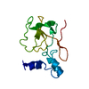
| ||||||||
|---|---|---|---|---|---|---|---|---|---|
| 1 |
| ||||||||
| Unit cell |
| ||||||||
| Atom site foot note | 1: RESIDUE PRO 95 IS A CIS PROLINE. |
-Components
| #1: Protein | Mass: 18024.672 Da / Num. of mol.: 1 Source method: isolated from a genetically manipulated source Source: (gene. exp.) |
|---|---|
| #2: Water | ChemComp-HOH /  Mass: 18.015 Da / Num. of mol.: 164 / Source method: isolated from a natural source / Formula: H2O Mass: 18.015 Da / Num. of mol.: 164 / Source method: isolated from a natural source / Formula: H2O |
| Has protein modification | Y |
-Experimental details
-Experiment
| Experiment | Method: X-RAY DIFFRACTION |
|---|
- Sample preparation
| Crystal | Density Matthews: 3.56 Å3/Da / Density % sol: 65.43 % | |||||||||||||||
|---|---|---|---|---|---|---|---|---|---|---|---|---|---|---|---|---|
| Crystal grow | *PLUS pH: 6.7 / Method: unknown | |||||||||||||||
| Components of the solutions | *PLUS
|
-Data collection
| Reflection | *PLUS Highest resolution: 2.25 Å / Num. all: 10599 / Num. obs: 8365 / % possible obs: 76 % / Observed criterion σ(F): 3 |
|---|
- Processing
| Software | Name: PROLSQ / Classification: refinement | ||||||||||||||||||||||||||||||||||||||||||||||||||||||||||||||||||||||||||||||||||||
|---|---|---|---|---|---|---|---|---|---|---|---|---|---|---|---|---|---|---|---|---|---|---|---|---|---|---|---|---|---|---|---|---|---|---|---|---|---|---|---|---|---|---|---|---|---|---|---|---|---|---|---|---|---|---|---|---|---|---|---|---|---|---|---|---|---|---|---|---|---|---|---|---|---|---|---|---|---|---|---|---|---|---|---|---|---|
| Refinement | Resolution: 2.8→5 Å / Rfactor obs: 0.175 / σ(F): 0 | ||||||||||||||||||||||||||||||||||||||||||||||||||||||||||||||||||||||||||||||||||||
| Refinement step | Cycle: LAST / Resolution: 2.8→5 Å
| ||||||||||||||||||||||||||||||||||||||||||||||||||||||||||||||||||||||||||||||||||||
| Refine LS restraints |
| ||||||||||||||||||||||||||||||||||||||||||||||||||||||||||||||||||||||||||||||||||||
| Software | *PLUS Name: PROLSQ / Classification: refinement | ||||||||||||||||||||||||||||||||||||||||||||||||||||||||||||||||||||||||||||||||||||
| Refinement | *PLUS Highest resolution: 2.25 Å / Lowest resolution: 5 Å / Num. reflection all: 6364 / σ(F): 0 / Rfactor all: 0.175 | ||||||||||||||||||||||||||||||||||||||||||||||||||||||||||||||||||||||||||||||||||||
| Solvent computation | *PLUS | ||||||||||||||||||||||||||||||||||||||||||||||||||||||||||||||||||||||||||||||||||||
| Displacement parameters | *PLUS |
