 Movie
Movie Controller

[English] 日本語
 Yorodumi
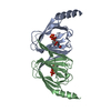
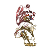
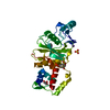
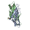
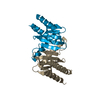
- PDB-2pak: Structure of a H51N mutant dTDP-4-keto-6-deoxy-D-glucose-3,4-keto... -
+ Open data

- Basic information
| Entry | Database: PDB / ID: 2pak | ||||||
|---|---|---|---|---|---|---|---|
| Title | Structure of a H51N mutant dTDP-4-keto-6-deoxy-D-glucose-3,4-ketoisomerase from Aneurinibacillus thermoaerophilus complexed with TDP | ||||||
 Components Components | DTDP-6-deoxy-3,4-keto-hexulose isomerase | ||||||
 Keywords Keywords | ISOMERASE / deoxysugar biosynthesis / S-Layer biosynthesis / ketoisomerase | ||||||
| Function / homology |  Function and homology information Function and homology informationTDP-4-oxo-6-deoxy-alpha-D-glucose-3,4-oxoisomerase (dTDP-3-dehydro-6-deoxy-alpha-D-galactopyranose-forming) / isomerase activity Similarity search - Function | ||||||
| Biological species |  Aneurinibacillus thermoaerophilus (bacteria) Aneurinibacillus thermoaerophilus (bacteria) | ||||||
| Method |  X-RAY DIFFRACTION / MOLECULAR REPLACEMENT / Resolution: 2.4 Å X-RAY DIFFRACTION / MOLECULAR REPLACEMENT / Resolution: 2.4 Å | ||||||
 Authors Authors | Davis, M.L. / Thoden, J.B. / Holden, H.M. | ||||||
 Citation Citation | Journal: J.Biol.Chem. / Year: 2007 Title: The X-ray Structure of dTDP-4-Keto-6-deoxy-D-glucose-3,4-ketoisomerase. Authors: Davis, M.L. / Thoden, J.B. / Holden, H.M. | ||||||
| History |
|
- Structure visualization
| Structure viewer | Molecule: MolmilJmol/JSmol |
|---|
- Downloads & links
-Download
| PDBx/mmCIF format | 2pak.cif.gz | 71.6 KB | Display | PDBx/mmCIF format |
|---|---|---|---|---|
| PDB format | pdb2pak.ent.gz | 52.8 KB | Display | PDB format |
| PDBx/mmJSON format | 2pak.json.gz | Tree view | PDBx/mmJSON format | |
| Others |  Other downloads Other downloads |
-Validation report
| Arichive directory | https://data.pdbj.org/pub/pdb/validation_reports/pa/2pakftp://data.pdbj.org/pub/pdb/validation_reports/pa/2pak | HTTPS FTP |
|---|
-Related structure data
| Related structure data | 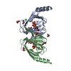 2pa7SC 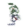 2paeC  2pamC S: Starting model for refinement C: citing same article ( |
|---|---|
| Similar structure data |


-Links
 PDBj
PDBj- Assembly
| Deposited unit | 
| ||||||||
|---|---|---|---|---|---|---|---|---|---|
| 1 |
| ||||||||
| Unit cell |
|
-Components
| #1: Protein | Mass: 16234.509 Da / Num. of mol.: 2 / Mutation: H51N Source method: isolated from a genetically manipulated source Source: (gene. exp.) Aneurinibacillus thermoaerophilus (bacteria)Strain: L420-91T / Gene: fdtA / Plasmid: pET28 / Production host: References: UniProt: Q6T1W8, Isomerases; Intramolecular oxidoreductases; Interconverting aldoses and ketoses, and related compounds #2: Chemical |   Mass: 402.188 Da / Num. of mol.: 2 / Source method: obtained synthetically / Formula: C10H16N2O11P2 Mass: 402.188 Da / Num. of mol.: 2 / Source method: obtained synthetically / Formula: C10H16N2O11P2#3: Water | ChemComp-HOH / |  Mass: 18.015 Da / Num. of mol.: 121 / Source method: isolated from a natural source / Formula: H2O Mass: 18.015 Da / Num. of mol.: 121 / Source method: isolated from a natural source / Formula: H2O |
|---|
-Experimental details
-Experiment
| Experiment | Method: X-RAY DIFFRACTION / Number of used crystals: 1 |
|---|
- Sample preparation
| Crystal | Density Matthews: 2.91 Å3/Da / Density % sol: 57.77 % |
|---|---|
| Crystal grow | Temperature: 298 K / Method: vapor diffusion, hanging drop / pH: 9 Details: 18% PEG 3400, 150mM LiCl, 100mM CHES, 10mM TDP, 20mM fucose, pH 9.0, VAPOR DIFFUSION, HANGING DROP, temperature 298K |
-Data collection
| Diffraction | Mean temperature: 100 K |
|---|---|
| Diffraction source | Source: ROTATING ANODE / Type: RIGAKU RU200 / Wavelength: 1.5418 |
| Detector | Type: Bruker Platinum 135 / Detector: CCD / Date: Mar 14, 2007 / Details: Montel |
| Radiation | Monochromator: nickel filter / Protocol: SINGLE WAVELENGTH / Monochromatic (M) / Laue (L): M / Scattering type: x-ray |
| Radiation wavelength | Wavelength: 1.5418 Å / Relative weight: 1 |
| Reflection | Resolution: 2.4→30 Å / Num. all: 15617 / Num. obs: 15617 / % possible obs: 97.5 % / Observed criterion σ(F): 0 / Observed criterion σ(I): 0 / Redundancy: 9.6 % / Rmerge(I) obs: 0.0959 / Rsym value: 0.0959 / Net I/σ(I): 15.7 |
| Reflection shell | Resolution: 2.39→2.49 Å / Redundancy: 3.1 % / Rmerge(I) obs: 0.422 / Mean I/σ(I) obs: 2.3 / Num. unique all: 1595 / Rsym value: 0.422 / % possible all: 90.7 |
- Processing
| Software |
| |||||||||||||||||||||||||
|---|---|---|---|---|---|---|---|---|---|---|---|---|---|---|---|---|---|---|---|---|---|---|---|---|---|---|
| Refinement | Method to determine structure: MOLECULAR REPLACEMENT Starting model: PDB ID 2PA7 Resolution: 2.4→30 Å / Isotropic thermal model: overall / Cross valid method: THROUGHOUT / σ(F): 0 / σ(I): 0 / Stereochemistry target values: Engh & Huber
| |||||||||||||||||||||||||
| Refinement step | Cycle: LAST / Resolution: 2.4→30 Å
|
