 Movie
Movie Controller

[English] 日本語
 Yorodumi
- PDB-2oxb: Crystal structure of a cell-wall invertase (E203Q) from Arabidops... -
+ Open data

- Basic information
| Entry | Database: PDB / ID: 2oxb | |||||||||
|---|---|---|---|---|---|---|---|---|---|---|
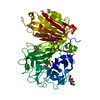
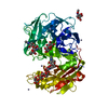
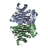
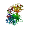
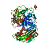
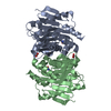
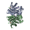
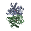
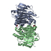
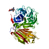
| Title | Crystal structure of a cell-wall invertase (E203Q) from Arabidopsis thaliana in complex with sucrose | |||||||||
 Components Components | Beta-fructofuranosidase | |||||||||
 Keywords Keywords | HYDROLASE | |||||||||
| Function / homology |  Function and homology information Function and homology informationbeta-fructofuranosidase activity / beta-fructofuranosidase / plant-type cell wall / apoplast / defense response to fungus / response to wounding / cytoplasmic stress granule / carbohydrate metabolic process / plasma membrane Similarity search - Function | |||||||||
| Biological species |  | |||||||||
| Method |  X-RAY DIFFRACTION / SYNCHROTRON / MOLECULAR REPLACEMENT / Resolution: 2.6 Å X-RAY DIFFRACTION / SYNCHROTRON / MOLECULAR REPLACEMENT / Resolution: 2.6 Å | |||||||||
 Authors Authors | Lammens, W. / Le Roy, K. / Van Laere, A. / Van den Ende, W. / Rabijns, A. | |||||||||
 Citation Citation | Journal: Proteins / Year: 2007 Title: An alternate sucrose binding mode in the E203Q Arabidopsis invertase mutant: An X-ray crystallography and docking study. Authors: Matrai, J. / Lammens, W. / Jonckheer, A. / Le Roy, K. / Rabijns, A. / Van den Ende, W. / De Maeyer, M. | |||||||||
| History |
|
- Structure visualization
| Structure viewer | Molecule: MolmilJmol/JSmol |
|---|
- Downloads & links
-Download
| PDBx/mmCIF format | 2oxb.cif.gz | 124.5 KB | Display | PDBx/mmCIF format |
|---|---|---|---|---|
| PDB format | pdb2oxb.ent.gz | 94.1 KB | Display | PDB format |
| PDBx/mmJSON format | 2oxb.json.gz | Tree view | PDBx/mmJSON format | |
| Others |  Other downloads Other downloads |
-Validation report
| Arichive directory | https://data.pdbj.org/pub/pdb/validation_reports/ox/2oxbftp://data.pdbj.org/pub/pdb/validation_reports/ox/2oxb | HTTPS FTP |
|---|
-Related structure data
| Related structure data |  2ac1S S: Starting model for refinement |
|---|---|
| Similar structure data |
-Links
 PDBj
PDBj- Assembly
| Deposited unit | 
| ||||||||
|---|---|---|---|---|---|---|---|---|---|
| 1 |
| ||||||||
| Unit cell |
|
-Components
| #1: Protein | Mass: 61102.055 Da / Num. of mol.: 1 / Mutation: E203Q Source method: isolated from a genetically manipulated source Source: (gene. exp.)  Pichia pastoris (fungus) / References: UniProt: Q43866, beta-fructofuranosidase Pichia pastoris (fungus) / References: UniProt: Q43866, beta-fructofuranosidase | ||||
|---|---|---|---|---|---|
| #2: Polysaccharide | beta-D-fructofuranose-(2-1)-alpha-D-glucopyranose / sucrose  Source method: isolated from a genetically manipulated source Details: oligosaccharide with reducing-end-to-reducing-end glycosidic bond References: sucrose | ||||
| #3: Sugar |   Type: D-saccharide, beta linking / Mass: 221.208 Da / Num. of mol.: 3 Type: D-saccharide, beta linking / Mass: 221.208 Da / Num. of mol.: 3Source method: isolated from a genetically manipulated source Formula: C8H15NO6 #4: Water | ChemComp-HOH / |  Mass: 18.015 Da / Num. of mol.: 34 / Source method: isolated from a natural source / Formula: H2O Mass: 18.015 Da / Num. of mol.: 34 / Source method: isolated from a natural source / Formula: H2OHas protein modification | Y | |
-Experimental details
-Experiment
| Experiment | Method: X-RAY DIFFRACTION / Number of used crystals: 1 |
|---|
- Sample preparation
| Crystal | Density Matthews: 2.7 Å3/Da / Density % sol: 54.52 % |
|---|---|
| Crystal grow | Temperature: 277 K / Method: vapor diffusion, hanging drop / pH: 6.5 Details: 25% PEG 4000, 0.1M zinc acetate, 0.1M sodium cacodylate, pH 6.5, VAPOR DIFFUSION, HANGING DROP, temperature 277K |
-Data collection
| Diffraction | Mean temperature: 100 K |
|---|---|
| Diffraction source | Source: SYNCHROTRON / Site: EMBL/DESY, HAMBURG  / Beamline: BW7A / Wavelength: 0.9837 / Beamline: BW7A / Wavelength: 0.9837 |
| Detector | Type: MAR CCD 165 mm / Detector: CCD / Date: Oct 26, 2005 |
| Radiation | Monochromator: Fixed exit double crystal Si [111], horizontally focussing Protocol: SINGLE WAVELENGTH / Monochromatic (M) / Laue (L): M / Scattering type: x-ray |
| Radiation wavelength | Wavelength: 0.9837 Å / Relative weight: 1 |
| Reflection | Resolution: 2.6→30 Å / Num. all: 19700 / Num. obs: 18370 / % possible obs: 99.7 % / Observed criterion σ(F): 2 / Observed criterion σ(I): 1.41 / Biso Wilson estimate: 50.9 Å2 |
| Reflection shell | Resolution: 2.6→2.64 Å / Rmerge(I) obs: 0.462 / Mean I/σ(I) obs: 2.29 / % possible all: 98 |
- Processing
| Software |
| ||||||||||||||||||||||||||||||||||||
|---|---|---|---|---|---|---|---|---|---|---|---|---|---|---|---|---|---|---|---|---|---|---|---|---|---|---|---|---|---|---|---|---|---|---|---|---|---|
| Refinement | Method to determine structure: MOLECULAR REPLACEMENT Starting model: PDB ENTRY 2AC1 Resolution: 2.6→19.51 Å / Rfactor Rfree error: 0.009 / Data cutoff high absF: 222814.77 / Data cutoff low absF: 0 / Isotropic thermal model: RESTRAINED / Cross valid method: THROUGHOUT / σ(F): 0 / Stereochemistry target values: Engh & Huber
| ||||||||||||||||||||||||||||||||||||
| Solvent computation | Solvent model: FLAT MODEL / Bsol: 53.7409 Å2 / ksol: 0.275024 e/Å3 | ||||||||||||||||||||||||||||||||||||
| Displacement parameters | Biso mean: 33.4 Å2 | ||||||||||||||||||||||||||||||||||||
| Refine analyze |
| ||||||||||||||||||||||||||||||||||||
| Refinement step | Cycle: LAST / Resolution: 2.6→19.51 Å
| ||||||||||||||||||||||||||||||||||||
| Refine LS restraints |
| ||||||||||||||||||||||||||||||||||||
| LS refinement shell | Resolution: 2.6→2.76 Å / Rfactor Rfree error: 0.029 / Total num. of bins used: 6
| ||||||||||||||||||||||||||||||||||||
| Xplor file |
|
