 Movie
Movie Controller


+ Open data

- Basic information
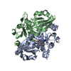
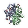
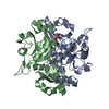
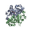
| Entry | Database: PDB / ID: 2owv | ||||||
|---|---|---|---|---|---|---|---|
| Title | Crystal structure of PH0725 from Pyrococcus horikoshii OT3 | ||||||
 Components Components | diphthine synthase | ||||||
 Keywords Keywords | TRANSFERASE / Pyrococcus horikoshii OT3 / Structural Genomics / NPPSFA / National Project on Protein Structural and Functional Analyses / RIKEN Structural Genomics/Proteomics Initiative / RSGI | ||||||
| Function / homology |  Function and homology information Function and homology informationdiphthine synthase / diphthine synthase activity / protein histidyl modification to diphthamide / methylation Similarity search - Function | ||||||
| Biological species |   Pyrococcus horikoshii (archaea) Pyrococcus horikoshii (archaea) | ||||||
| Method |  X-RAY DIFFRACTION / FOURIER SYNTHESIS / Resolution: 2.8 Å X-RAY DIFFRACTION / FOURIER SYNTHESIS / Resolution: 2.8 Å | ||||||
 Authors Authors | Sugahara, M. / Kageyama, Y. / Matsuura, Y. / Shimada, H. / Kunishima, N. / RIKEN Structural Genomics/Proteomics Initiative (RSGI) | ||||||
 Citation Citation | Journal: To be Published Title: Crystal structure of PH0725 from Pyrococcus horikoshii OT3 Authors: Sugahara, M. / Kageyama, Y. / Matsuura, Y. / Shimada, H. / Kunishima, N. | ||||||
| History |
|
- Structure visualization
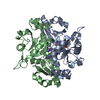
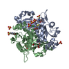
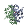
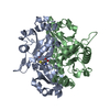
| Structure viewer | Molecule: MolmilJmol/JSmol |
|---|
- Downloads & links
-Download
| PDBx/mmCIF format | 2owv.cif.gz | 117.2 KB | Display | PDBx/mmCIF format |
|---|---|---|---|---|
| PDB format | pdb2owv.ent.gz | 91.1 KB | Display | PDB format |
| PDBx/mmJSON format | 2owv.json.gz | Tree view | PDBx/mmJSON format | |
| Others |  Other downloads Other downloads |
-Validation report
| Arichive directory | https://data.pdbj.org/pub/pdb/validation_reports/ow/2owvftp://data.pdbj.org/pub/pdb/validation_reports/ow/2owv | HTTPS FTP |
|---|
-Related structure data
| Related structure data |  2e8hC 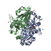 2hr8C  2huqC 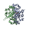 2hutC 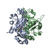 2huvC 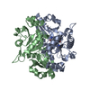 2huxC 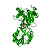 2owfC  2owgC  2owkC 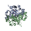 2owuC 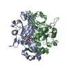 2p2xC 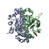 2p5cC 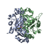 2p5fC  2p6dC 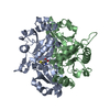 2p6iC 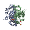 2p6kC 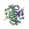 2p6lC 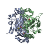 2p9dC 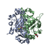 2pb4C 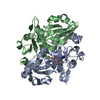 2pb5C 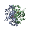 2pb6C 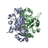 2pcaC 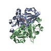 2pcgC 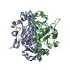 2pchC 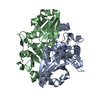 2pciC 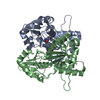 2pckC 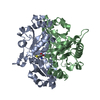 2pcmC 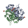 1wngS S: Starting model for refinement C: citing same article ( |
|---|---|
| Similar structure data | |
| Other databases |


-Links
 PDBj
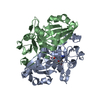
PDBj- Assembly
| Deposited unit | 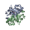
| ||||||||
|---|---|---|---|---|---|---|---|---|---|
| 1 |
| ||||||||
| 2 | 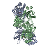
| ||||||||
| Unit cell |
| ||||||||
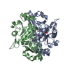
| Details | The biological assembly is a dimer. |
-Components
| #1: Protein | Mass: 29581.389 Da / Num. of mol.: 2 / Mutation: Y148M Source method: isolated from a genetically manipulated source Source: (gene. exp.) Pyrococcus horikoshii (archaea) / Strain: OT3 / Plasmid: pET-11A / Production host:  #2: Chemical | ChemComp-SAH / |   Type: L-peptide linking / Mass: 384.411 Da / Num. of mol.: 1 / Source method: obtained synthetically / Formula: C14H20N6O5S Type: L-peptide linking / Mass: 384.411 Da / Num. of mol.: 1 / Source method: obtained synthetically / Formula: C14H20N6O5S#3: Water | ChemComp-HOH / |  Mass: 18.015 Da / Num. of mol.: 121 / Source method: isolated from a natural source / Formula: H2O Mass: 18.015 Da / Num. of mol.: 121 / Source method: isolated from a natural source / Formula: H2O |
|---|
-Experimental details
-Experiment
| Experiment | Method: X-RAY DIFFRACTION / Number of used crystals: 1 |
|---|
- Sample preparation
| Crystal | Density Matthews: 3.28 Å3/Da / Density % sol: 62.48 % |
|---|---|
| Crystal grow | Temperature: 295 K / Method: oil microbatch / pH: 5.5 Details: 3.86M Sodium formate, 0.1M Acetate, pH 5.5, oil microbatch, temperature 295K |
-Data collection
| Diffraction | Mean temperature: 100 K |
|---|---|
| Diffraction source | Source: ROTATING ANODE / Type: RIGAKU / Wavelength: 1.54 Å |
| Detector | Type: RIGAKU RAXIS V / Detector: IMAGE PLATE / Date: Sep 6, 2006 |
| Radiation | Protocol: SINGLE WAVELENGTH / Monochromatic (M) / Laue (L): M / Scattering type: x-ray |
| Radiation wavelength | Wavelength: 1.54 Å / Relative weight: 1 |
| Reflection | Resolution: 2.8→15 Å / Num. all: 19853 / Num. obs: 19853 / % possible obs: 99.8 % / Observed criterion σ(F): 0 / Observed criterion σ(I): 0 / Redundancy: 9.2 % / Biso Wilson estimate: 75.8 Å2 / Rmerge(I) obs: 0.095 / Rsym value: 0.092 / Net I/σ(I): 8.6 |
| Reflection shell | Resolution: 2.8→2.9 Å / Redundancy: 9.5 % / Rmerge(I) obs: 0.586 / Mean I/σ(I) obs: 3.6 / Num. unique all: 1934 / Rsym value: 0.56 / % possible all: 100 |
- Processing
| Software |
| |||||||||||||||||||||||||
|---|---|---|---|---|---|---|---|---|---|---|---|---|---|---|---|---|---|---|---|---|---|---|---|---|---|---|
| Refinement | Method to determine structure: FOURIER SYNTHESIS Starting model: PDB ENTRY 1wng Resolution: 2.8→14.97 Å / Isotropic thermal model: Anisotrop / Cross valid method: THROUGHOUT / σ(F): 0 / Stereochemistry target values: Engh & Huber
| |||||||||||||||||||||||||
| Displacement parameters | Biso mean: 54.8 Å2
| |||||||||||||||||||||||||
| Refine analyze |
| |||||||||||||||||||||||||
| Refinement step | Cycle: LAST / Resolution: 2.8→14.97 Å
| |||||||||||||||||||||||||
| Refine LS restraints |
| |||||||||||||||||||||||||
| LS refinement shell | Resolution: 2.8→2.9 Å / Rfactor Rfree error: 0.038
|
