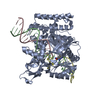
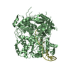
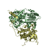
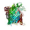
DNA ligase (NAD+) / DNA ligase (NAD+) activity / NAD+ binding / DNA replication / DNA repair / DNA binding / metal ion binding / cytosol Similarity search - Function
Laminin - #30 / Laminin / Dna Ligase; domain 1 - #70 / Zinc-finger, NAD-dependent DNA ligase C4-type / NAD-dependent DNA ligase C4 zinc finger domain / NAD-dependent DNA ligase, active site / NAD-dependent DNA ligase, conserved site / NAD-dependent DNA ligase signature 1. / NAD-dependent DNA ligase signature 2. / NAD-dependent DNA ligase ...Laminin - #30 / Laminin / Dna Ligase; domain 1 - #70 / Zinc-finger, NAD-dependent DNA ligase C4-type / NAD-dependent DNA ligase C4 zinc finger domain / NAD-dependent DNA ligase, active site / NAD-dependent DNA ligase, conserved site / NAD-dependent DNA ligase signature 1. / NAD-dependent DNA ligase signature 2. / NAD-dependent DNA ligase / NAD-dependent DNA ligase, OB-fold / NAD-dependent DNA ligase, adenylation / NAD-dependent DNA ligase, N-terminal / NAD-dependent DNA ligase adenylation domain / NAD-dependent DNA ligase OB-fold domain / Ligase N family / DisA/LigA, helix-hairpin-helix motif / Helix-hairpin-helix motif / DNA ligase/mRNA capping enzyme / Helix hairpin bin / RuvA domain 2-like / Other non-globular / BRCA1 C Terminus (BRCT) domain / Helix-hairpin-helix domain / breast cancer carboxy-terminal domain / D-amino Acid Aminotransferase; Chain A, domain 1 / Helix-hairpin-helix DNA-binding motif, class 1 / Helix-hairpin-helix DNA-binding motif class 1 / BRCT domain profile. / BRCT domain / BRCT domain superfamily / Nucleic acid-binding proteins / Dna Ligase; domain 1 / 5' to 3' exonuclease, C-terminal subdomain / Special / DNA polymerase; domain 1 / OB fold (Dihydrolipoamide Acetyltransferase, E2P) / Helix Hairpins / Nucleic acid-binding, OB-fold / Beta Barrel / 2-Layer Sandwich / Orthogonal Bundle / Mainly Beta / Mainly Alpha / Alpha Beta Similarity search - Domain/homology
Mass: 18.015 Da / Num. of mol.: 320 / Source method: isolated from a natural source / Formula: H2O
-
Experimental details
-
Experiment
Experiment
Method: X-RAY DIFFRACTION / Number of used crystals: 1
-
Sample preparation
Crystal
Density Matthews: 2.52 Å3/Da / Density % sol: 51.28 %
Crystal grow
Temperature: 295 K / Method: vapor diffusion, sitting drop / pH: 6 Details: EcoLigA (0.3 mM) was reacted with 5 mM MgCl2 and 0.3 mM NAD+ for 30 min at 22 C. The ligase-adenylylation reaction was then quenched by adding 10 mM EDTA. The mixture was supplemented with ...Details: EcoLigA (0.3 mM) was reacted with 5 mM MgCl2 and 0.3 mM NAD+ for 30 min at 22 C. The ligase-adenylylation reaction was then quenched by adding 10 mM EDTA. The mixture was supplemented with 26-bp nicked duplex DNA (0.318 mM). This LigA-nucleic acid solution was mixed 1:2 with a well solution containing 200 mM ammonium sulfate, 50 mM sodium acetate, 24% PEG-4000. Crystals were grown at 22 C by the sitting drop vapor diffusion method. Crystals appeared after 3 days. The crystals were transferred serially to solutions containing 5% glycerol/24% PEG-4000, 10% glycerol/26% PEG-4000, 15% glycerol/28% PEG-4000, and 20% glycerol/30% PEG-4000 in 100 mM ammonium sulfate, 50 mM sodium acetate, after which they were flash-frozen in liquid nitrogen. , pH 6.0, VAPOR DIFFUSION, SITTING DROP, temperature 295K
In the structure databanks used in Yorodumi, some data are registered as the other names, "COVID-19 virus" and "2019-nCoV". Here are the details of the virus and the list of structure data.
Jan 31, 2019. EMDB accession codes are about to change! (news from PDBe EMDB page)
EMDB accession codes are about to change! (news from PDBe EMDB page)
The allocation of 4 digits for EMDB accession codes will soon come to an end. Whilst these codes will remain in use, new EMDB accession codes will include an additional digit and will expand incrementally as the available range of codes is exhausted. The current 4-digit format prefixed with “EMD-” (i.e. EMD-XXXX) will advance to a 5-digit format (i.e. EMD-XXXXX), and so on. It is currently estimated that the 4-digit codes will be depleted around Spring 2019, at which point the 5-digit format will come into force.
The EM Navigator/Yorodumi systems omit the EMD- prefix.
Related info.:Q: What is EMD? / ID/Accession-code notation in Yorodumi/EM Navigator
Yorodumi is a browser for structure data from EMDB, PDB, SASBDB, etc.
This page is also the successor to EM Navigator detail page, and also detail information page/front-end page for Omokage search.
The word "yorodu" (or yorozu) is an old Japanese word meaning "ten thousand". "mi" (miru) is to see.
Related info.:EMDB / PDB / SASBDB / Comparison of 3 databanks / Yorodumi Search / Aug 31, 2016. New EM Navigator & Yorodumi / Yorodumi Papers / Jmol/JSmol / Function and homology information / Changes in new EM Navigator and Yorodumi
 Movie
Movie Controller

 Yorodumi
 Open data
 Basic information
 Components
 Keywords
 Function and homology information
 X-RAY DIFFRACTION /
  Authors
 Citation
 Structure visualization
 Downloads & links
 Other downloads
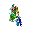


 PDBj
PDBj


 Assembly
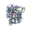


 Mass: 347.221 Da / Num. of mol.: 1 / Source method: obtained synthetically / Formula: C10H14N5O7P / Comment: AMP*YM
 Mass: 65.409 Da / Num. of mol.: 1 / Source method: obtained synthetically / Formula: Zn
 Mass: 96.063 Da / Num. of mol.: 4 / Source method: obtained synthetically / Formula: SO4
 Sample preparation
 / Beamline: 24-ID-C / Wavelength: 0.9792 Å
 Processing