- PDB-2ou6: Crystal structure of a putative metalloenzyme of the duf664 famil... -
+
Open data
ID or keywords:
Loading...
-
Basic information
Entry
Database: PDB / ID: 2ou6
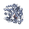
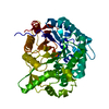
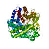
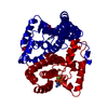
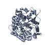
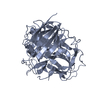
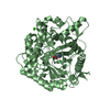
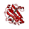
Title
Crystal structure of a putative metalloenzyme of the duf664 family (dr_1065) from deinococcus radiodurans at 1.80 A resolution
Components
Hypothetical protein
Keywords
METAL BINDING PROTEIN / Structural genomics / Joint Center for Structural Genomics / JCSG / Protein Structure Initiative / PSI-2
Function / homology
Protein of unknown function DUF664 / Mycothiol-S-transferase / dinb family like domain / DinB/YfiT-like putative metalloenzymes / Four Helix Bundle (Hemerythrin (Met), subunit A) / Up-down Bundle / Mainly Alpha / NICKEL (II) ION / DinB-like domain-containing protein
Function and homology information
Biological species
Deinococcus radiodurans (radioresistant)
Method
X-RAY DIFFRACTION / SYNCHROTRON / MAD / Resolution: 1.8 Å
SEQUENCE THE CONSTRUCT WAS EXPRESSED WITH A PURIFICATION TAG MGSDKIHHHHHHENLYFQG. THE TAG WAS ... SEQUENCE THE CONSTRUCT WAS EXPRESSED WITH A PURIFICATION TAG MGSDKIHHHHHHENLYFQG. THE TAG WAS REMOVED WITH TEV PROTEASE LEAVING ONLY A GLYCINE, FOLLOWED BY THE TARGET SEQUENCE.
Type: MARMOSAIC 325 mm CCD / Detector: CCD / Date: Feb 1, 2007 / Details: Flat mirror (vertical focusing)
Radiation
Monochromator: Single crystal Si(111) bent (horizontal focusing) Protocol: MAD / Monochromatic (M) / Laue (L): M / Scattering type: x-ray
Radiation wavelength
ID
Wavelength (Å)
Relative weight
1
0.97905
1
2
0.97932
1
3
0.91837
1
Reflection
Resolution: 1.8→125 Å / Num. obs: 22207 / % possible obs: 91.1 % / Redundancy: 6.9 % / Biso Wilson estimate: 21.72 Å2 / Rmerge(I) obs: 0.07 / Rsym value: 0.07 / Net I/σ(I): 7.1
Reflection shell
Diffraction-ID: 1
Resolution (Å)
Redundancy (%)
Rmerge(I) obs
Mean I/σ(I) obs
Num. measured all
Num. unique all
Rsym value
% possible all
1.8-1.85
2.6
0.268
2.5
2678
1047
0.268
61.4
1.85-1.9
3
0.237
2.7
3766
1244
0.237
74.1
1.9-1.95
3.8
0.219
2.8
5100
1340
0.219
82
1.95-2.01
5.2
0.203
3.1
7047
1364
0.203
83.7
2.01-2.08
5.1
0.16
3.9
7020
1372
0.16
87.5
2.08-2.15
5
0.133
4.5
6977
1383
0.133
92
2.15-2.23
5.2
0.119
5.1
7278
1404
0.119
94.9
2.23-2.32
5.3
0.108
5.4
7344
1383
0.108
98.4
2.32-2.43
5.6
0.096
6.2
7663
1362
0.096
99.6
2.43-2.55
6
0.095
6.3
7769
1293
0.095
99.9
2.55-2.68
6.6
0.093
6.4
8380
1262
0.093
100
2.68-2.85
7.3
0.086
7
8669
1192
0.086
100
2.85-3.04
7.9
0.077
7.8
9090
1145
0.077
100
3.04-3.29
8.8
0.066
8.9
9158
1043
0.066
100
3.29-3.6
10.2
0.062
9.6
10158
999
0.062
100
3.6-4.02
12.6
0.062
9.3
11380
901
0.062
100
4.02-4.65
14.4
0.061
10.2
11680
809
0.061
100
4.65-5.69
14
0.061
9.7
9936
711
0.061
100
5.69-8.05
13.3
0.066
8.6
7754
581
0.066
100
8.05-125.43
10.3
0.058
10.2
3843
372
0.058
98.3
-
Phasing
Phasing
Method: MAD
-
Processing
Software
Name
Version
Classification
NB
MolProbity
3beta29
modelbuilding
SHELX
phasing
REFMAC
5.2.0005
refinement
SCALA
datascaling
PDB_EXTRACT
2
dataextraction
MAR345
CCD
datacollection
MOSFLM
datareduction
CCP4
(SCALA)
datascaling
SHELXD
phasing
autoSHARP
phasing
Refinement
Method to determine structure: MAD / Resolution: 1.8→125 Å / Cor.coef. Fo:Fc: 0.962 / Cor.coef. Fo:Fc free: 0.944 / SU B: 3.232 / SU ML: 0.056 / TLS residual ADP flag: LIKELY RESIDUAL / Cross valid method: THROUGHOUT / σ(F): 0 / ESU R: 0.103 / ESU R Free: 0.103 Stereochemistry target values: MAXIMUM LIKELIHOOD WITH PHASES Details: 1. HYDROGENS HAVE BEEN ADDED IN THE RIDING POSITIONS. 2. A MET-INHIBITION PROTOCOL WAS USED FOR SELENOMETHIONINE INCORPORATION DURING PROTEIN EXPRESSION. THE OCCUPANCY OF THE SE ATOMS IN THE ...Details: 1. HYDROGENS HAVE BEEN ADDED IN THE RIDING POSITIONS. 2. A MET-INHIBITION PROTOCOL WAS USED FOR SELENOMETHIONINE INCORPORATION DURING PROTEIN EXPRESSION. THE OCCUPANCY OF THE SE ATOMS IN THE MSE RESIDUES WAS REDUCED TO 0.75 FOR THE REDUCED SCATTERING POWER DUE TO PARTIAL S-MET INCORPORATION. 3. ATOM RECORD CONTAINS RESIDUAL B FACTORS ONLY. 4. NI MODELED BASED ON GEOMETRY AND HOMOLOGS. 5. SO4 AND GOL MODELED BASED ON CRYSTALLIZATION CONDITIONS. 6. THERE IS UNMODELED DENSITY NEAR PHE 120 AND TRP 160.
Rfactor
Num. reflection
% reflection
Selection details
Rfree
0.188
1137
5.1 %
RANDOM
Rwork
0.155
-
-
-
all
0.157
-
-
-
obs
0.157
22101
91.06 %
-
Solvent computation
Ion probe radii: 0.8 Å / Shrinkage radii: 0.8 Å / VDW probe radii: 1.2 Å / Solvent model: MASK
Displacement parameters
Biso mean: 19.024 Å2
Baniso -1
Baniso -2
Baniso -3
1-
-1.32 Å2
-0.66 Å2
0 Å2
2-
-
-1.32 Å2
0 Å2
3-
-
-
1.98 Å2
Refinement step
Cycle: LAST / Resolution: 1.8→125 Å
Protein
Nucleic acid
Ligand
Solvent
Total
Num. atoms
1428
0
23
266
1717
Refine LS restraints
Refine-ID
Type
Dev ideal
Dev ideal target
Number
X-RAY DIFFRACTION
r_bond_refined_d
0.015
0.022
1517
X-RAY DIFFRACTION
r_bond_other_d
0.001
0.02
1354
X-RAY DIFFRACTION
r_angle_refined_deg
1.548
1.947
2083
X-RAY DIFFRACTION
r_angle_other_deg
0.912
3
3130
X-RAY DIFFRACTION
r_dihedral_angle_1_deg
5.626
5
192
X-RAY DIFFRACTION
r_dihedral_angle_2_deg
36.358
23.867
75
X-RAY DIFFRACTION
r_dihedral_angle_3_deg
11.829
15
217
X-RAY DIFFRACTION
r_dihedral_angle_4_deg
15.484
15
11
X-RAY DIFFRACTION
r_chiral_restr
0.102
0.2
227
X-RAY DIFFRACTION
r_gen_planes_refined
0.008
0.02
1718
X-RAY DIFFRACTION
r_gen_planes_other
0.001
0.02
315
X-RAY DIFFRACTION
r_nbd_refined
0.217
0.2
322
X-RAY DIFFRACTION
r_nbd_other
0.185
0.2
1293
X-RAY DIFFRACTION
r_nbtor_refined
0.181
0.2
754
X-RAY DIFFRACTION
r_nbtor_other
0.087
0.2
805
X-RAY DIFFRACTION
r_xyhbond_nbd_refined
0.161
0.2
201
X-RAY DIFFRACTION
r_symmetry_vdw_refined
0.136
0.2
12
X-RAY DIFFRACTION
r_symmetry_vdw_other
0.303
0.2
49
X-RAY DIFFRACTION
r_symmetry_hbond_refined
0.089
0.2
11
X-RAY DIFFRACTION
r_mcbond_it
2.02
3
998
X-RAY DIFFRACTION
r_mcbond_other
0.505
3
373
X-RAY DIFFRACTION
r_mcangle_it
2.609
5
1489
X-RAY DIFFRACTION
r_scbond_it
4.621
8
653
X-RAY DIFFRACTION
r_scangle_it
6.492
11
589
LS refinement shell
Resolution: 1.8→1.847 Å / Total num. of bins used: 20
Rfactor
Num. reflection
% reflection
Rfree
0.251
73
-
Rwork
0.175
973
-
obs
-
1046
60.71 %
Refinement TLS params.
Method: refined / Origin x: 13.647 Å / Origin y: 39.193 Å / Origin z: 2.493 Å
11
12
13
21
22
23
31
32
33
T
0.0107 Å2
-0.0021 Å2
0.0017 Å2
-
-0.0383 Å2
-0.0119 Å2
-
-
-0.089 Å2
L
0.4919 °2
0.1147 °2
-0.0674 °2
-
0.7817 °2
-0.0598 °2
-
-
0.5193 °2
S
0.0203 Å °
-0.0194 Å °
-0.0413 Å °
0.0356 Å °
-0.0112 Å °
0.0191 Å °
0.0985 Å °
-0.0105 Å °
-0.0091 Å °
Refinement TLS group
Selection: ALL
+
About Yorodumi
-
News
-
Feb 9, 2022. New format data for meta-information of EMDB entries
New format data for meta-information of EMDB entries
Version 3 of the EMDB header file is now the official format.
The previous official version 1.9 will be removed from the archive.
In the structure databanks used in Yorodumi, some data are registered as the other names, "COVID-19 virus" and "2019-nCoV". Here are the details of the virus and the list of structure data.
Jan 31, 2019. EMDB accession codes are about to change! (news from PDBe EMDB page)
EMDB accession codes are about to change! (news from PDBe EMDB page)
The allocation of 4 digits for EMDB accession codes will soon come to an end. Whilst these codes will remain in use, new EMDB accession codes will include an additional digit and will expand incrementally as the available range of codes is exhausted. The current 4-digit format prefixed with “EMD-” (i.e. EMD-XXXX) will advance to a 5-digit format (i.e. EMD-XXXXX), and so on. It is currently estimated that the 4-digit codes will be depleted around Spring 2019, at which point the 5-digit format will come into force.
The EM Navigator/Yorodumi systems omit the EMD- prefix.
Related info.:Q: What is EMD? / ID/Accession-code notation in Yorodumi/EM Navigator
Yorodumi is a browser for structure data from EMDB, PDB, SASBDB, etc.
This page is also the successor to EM Navigator detail page, and also detail information page/front-end page for Omokage search.
The word "yorodu" (or yorozu) is an old Japanese word meaning "ten thousand". "mi" (miru) is to see.
Related info.:EMDB / PDB / SASBDB / Comparison of 3 databanks / Yorodumi Search / Aug 31, 2016. New EM Navigator & Yorodumi / Yorodumi Papers / Jmol/JSmol / Function and homology information / Changes in new EM Navigator and Yorodumi
 Movie
Movie Controller

 Yorodumi
 Open data
 Basic information
 Components
 Keywords
 Function and homology information
 Deinococcus radiodurans (radioresistant)
 X-RAY DIFFRACTION /
  Authors
 Citation
 Structure visualization
 Downloads & links
 Other downloads


 PDBj
PDBj Assembly
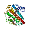

 Mass: 58.693 Da / Num. of mol.: 1 / Source method: obtained synthetically / Formula: Ni
 Mass: 96.063 Da / Num. of mol.: 2 / Source method: obtained synthetically / Formula: SO4
 Mass: 92.094 Da / Num. of mol.: 2 / Source method: obtained synthetically / Formula: C3H8O3
 Mass: 18.015 Da / Num. of mol.: 266 / Source method: isolated from a natural source / Formula: H2O
 Sample preparation
 / Beamline: BL11-1 / Wavelength: 0.97905, 0.97932, 0.91837
 Processing