SEQUENCE THE CONSTRUCT WAS EXPRESSED WITH A PURIFICATION TAG MGSDKIHHHHHHENLYFQG. THE TAG WAS ... SEQUENCE THE CONSTRUCT WAS EXPRESSED WITH A PURIFICATION TAG MGSDKIHHHHHHENLYFQG. THE TAG WAS REMOVED WITH TEV PROTEASE LEAVING ONLY A GLYCINE, FOLLOWED BY THE TARGET SEQUENCE.
Type: MARMOSAIC 325 mm CCD / Detector: CCD / Date: Jan 31, 2007 / Details: Flat mirror (vertical focusing)
Radiation
Monochromator: Single crystal Si(111) bent (horizontal focusing) Protocol: MAD / Monochromatic (M) / Laue (L): M / Scattering type: x-ray
Radiation wavelength
ID
Wavelength (Å)
Relative weight
1
0.91837
1
2
0.97931
1
3
0.97885
1
Reflection
Resolution: 1.85→29.21 Å / Num. obs: 46806 / % possible obs: 100 % / Redundancy: 3.8 % / Biso Wilson estimate: 19.11 Å2 / Rmerge(I) obs: 0.105 / Rsym value: 0.105 / Net I/σ(I): 5.9
Reflection shell
Diffraction-ID: 1
Resolution (Å)
Redundancy (%)
Rmerge(I) obs
Mean I/σ(I) obs
Num. measured all
Num. unique all
Rsym value
% possible all
1.85-1.9
3.8
0.667
1.1
13148
3463
0.667
100
1.9-1.95
3.8
0.522
1.4
12642
3338
0.522
99.8
1.95-2.01
3.8
0.423
1.8
12361
3259
0.423
100
2.01-2.07
3.8
0.341
2.2
12068
3177
0.341
100
2.07-2.14
3.8
0.274
2.7
11725
3086
0.274
100
2.14-2.21
3.8
0.246
3
11271
2960
0.246
100
2.21-2.29
3.8
0.219
3.4
10946
2880
0.219
100
2.29-2.39
3.8
0.195
3.8
10498
2757
0.195
100
2.39-2.49
3.8
0.162
4.6
10086
2648
0.162
100
2.49-2.62
3.8
0.145
5.2
9743
2556
0.145
100
2.62-2.76
3.8
0.119
6
9127
2391
0.119
100
2.76-2.93
3.8
0.098
7.4
8834
2316
0.098
100
2.93-3.13
3.8
0.085
8
8147
2135
0.085
100
3.13-3.38
3.8
0.069
9.6
7704
2020
0.069
100
3.38-3.7
3.8
0.059
10.7
7063
1857
0.059
100
3.7-4.14
3.8
0.052
11.4
6373
1674
0.052
100
4.14-4.78
3.8
0.049
12.2
5621
1482
0.049
100
4.78-5.85
3.8
0.052
10.8
4824
1278
0.052
100
5.85-8.27
3.7
0.053
11.5
3651
981
0.053
100
8.27-29.21
3.6
0.052
10.8
1958
548
0.052
97.8
-
Phasing
Phasing
Method: MAD
-
Processing
Software
Name
Version
Classification
NB
MolProbity
3beta29
modelbuilding
SHELX
phasing
REFMAC
5.2.0019
refinement
SCALA
datascaling
PDB_EXTRACT
2
dataextraction
MAR345
CCD
datacollection
MOSFLM
datareduction
CCP4
(SCALA)
datascaling
SHELXD
phasing
autoSHARP
phasing
Refinement
Method to determine structure: MAD / Resolution: 1.85→29.21 Å / Cor.coef. Fo:Fc: 0.97 / Cor.coef. Fo:Fc free: 0.963 / SU B: 4.772 / SU ML: 0.072 / TLS residual ADP flag: LIKELY RESIDUAL / Cross valid method: THROUGHOUT / σ(F): 0 / ESU R: 0.106 / ESU R Free: 0.1 Stereochemistry target values: MAXIMUM LIKELIHOOD WITH PHASES Details: 1. HYDROGENS HAVE BEEN ADDED IN THE RIDING POSITIONS. 2. ATOM RECORD CONTAINS RESIDUAL B FACTORS ONLY. 3. A MET-INHIBITION PROTOCOL WAS USED FOR SELENOMETHIONINE INCORPORATION DURING PROTEIN ...Details: 1. HYDROGENS HAVE BEEN ADDED IN THE RIDING POSITIONS. 2. ATOM RECORD CONTAINS RESIDUAL B FACTORS ONLY. 3. A MET-INHIBITION PROTOCOL WAS USED FOR SELENOMETHIONINE INCORPORATION DURING PROTEIN EXPRESSION. THE OCCUPANCY OF THE SE ATOMS IN THE MSE RESIDUES WAS REDUCED TO 0.75 FOR THE REDUCED SCATTERING POWER DUE TO PARTIAL S-MET INCORPORATION. 4. CALCIUM, SODIUM, GLYCEROL AND ACETATE IONS ARE MODELED BASED ON THE CRYSTALLIZATION CONDITIONS AND ELECTRON DENSITY.
Rfactor
Num. reflection
% reflection
Selection details
Rfree
0.177
2366
5.1 %
RANDOM
Rwork
0.151
-
-
-
all
0.152
-
-
-
obs
0.152
46806
99.95 %
-
Solvent computation
Ion probe radii: 0.8 Å / Shrinkage radii: 0.8 Å / VDW probe radii: 1.2 Å / Solvent model: BABINET MODEL WITH MASK
Displacement parameters
Biso mean: 19.687 Å2
Baniso -1
Baniso -2
Baniso -3
1-
0.21 Å2
0 Å2
-0.17 Å2
2-
-
1.02 Å2
0 Å2
3-
-
-
-1.25 Å2
Refinement step
Cycle: LAST / Resolution: 1.85→29.21 Å
Protein
Nucleic acid
Ligand
Solvent
Total
Num. atoms
3448
0
34
426
3908
Refine LS restraints
Refine-ID
Type
Dev ideal
Dev ideal target
Number
X-RAY DIFFRACTION
r_bond_refined_d
0.015
0.022
3607
X-RAY DIFFRACTION
r_bond_other_d
0.002
0.02
2383
X-RAY DIFFRACTION
r_angle_refined_deg
1.484
1.991
4909
X-RAY DIFFRACTION
r_angle_other_deg
0.96
3
5849
X-RAY DIFFRACTION
r_dihedral_angle_1_deg
6.259
5
475
X-RAY DIFFRACTION
r_dihedral_angle_2_deg
34.977
24.178
146
X-RAY DIFFRACTION
r_dihedral_angle_3_deg
12.795
15
595
X-RAY DIFFRACTION
r_dihedral_angle_4_deg
15.382
15
20
X-RAY DIFFRACTION
r_chiral_restr
0.091
0.2
561
X-RAY DIFFRACTION
r_gen_planes_refined
0.006
0.02
4068
X-RAY DIFFRACTION
r_gen_planes_other
0.002
0.02
697
X-RAY DIFFRACTION
r_nbd_refined
0.204
0.2
731
X-RAY DIFFRACTION
r_nbd_other
0.2
0.2
2574
X-RAY DIFFRACTION
r_nbtor_refined
0.177
0.2
1764
X-RAY DIFFRACTION
r_nbtor_other
0.088
0.2
1981
X-RAY DIFFRACTION
r_xyhbond_nbd_refined
0.164
0.2
332
X-RAY DIFFRACTION
r_metal_ion_refined
0.17
0.2
13
X-RAY DIFFRACTION
r_symmetry_vdw_refined
0.737
0.2
9
X-RAY DIFFRACTION
r_symmetry_vdw_other
0.343
0.2
13
X-RAY DIFFRACTION
r_symmetry_hbond_refined
0.159
0.2
13
X-RAY DIFFRACTION
r_symmetry_metal_ion_refined
0.251
0.2
6
X-RAY DIFFRACTION
r_mcbond_it
1.394
3
2313
X-RAY DIFFRACTION
r_mcbond_other
0.358
3
938
X-RAY DIFFRACTION
r_mcangle_it
2.49
5
3680
X-RAY DIFFRACTION
r_scbond_it
4.462
8
1329
X-RAY DIFFRACTION
r_scangle_it
6.541
11
1218
Refine LS restraints NCS
Ens-ID: 1 / Refine-ID: X-RAY DIFFRACTION
Dom-ID
Auth asym-ID
Number
Type
Rms dev position (Å)
Weight position
1
A
847
TIGHTPOSITIONAL
0.08
0.05
2
B
847
TIGHTPOSITIONAL
0.04
0.05
3
C
847
TIGHTPOSITIONAL
0.07
0.05
1
A
924
MEDIUMPOSITIONAL
0.39
0.5
2
B
924
MEDIUMPOSITIONAL
0.34
0.5
3
C
924
MEDIUMPOSITIONAL
0.34
0.5
1
A
43
LOOSEPOSITIONAL
0.68
5
2
B
43
LOOSEPOSITIONAL
0.57
5
3
C
43
LOOSEPOSITIONAL
0.68
5
1
A
847
TIGHTTHERMAL
0.19
0.5
2
B
847
TIGHTTHERMAL
0.18
0.5
3
C
847
TIGHTTHERMAL
0.19
0.5
1
A
924
MEDIUMTHERMAL
0.86
2
2
B
924
MEDIUMTHERMAL
0.9
2
3
C
924
MEDIUMTHERMAL
0.9
2
1
A
43
LOOSETHERMAL
1.61
10
2
B
43
LOOSETHERMAL
4.59
10
3
C
43
LOOSETHERMAL
5.75
10
LS refinement shell
Resolution: 1.85→1.898 Å / Total num. of bins used: 20
Rfactor
Num. reflection
% reflection
Rfree
0.268
160
-
Rwork
0.226
3301
-
obs
-
3461
99.97 %
Refinement TLS params.
Method: refined / Refine-ID: X-RAY DIFFRACTION
ID
L11 (°2)
L12 (°2)
L13 (°2)
L22 (°2)
L23 (°2)
L33 (°2)
S11 (Å °)
S12 (Å °)
S13 (Å °)
S21 (Å °)
S22 (Å °)
S23 (Å °)
S31 (Å °)
S32 (Å °)
S33 (Å °)
T11 (Å2)
T12 (Å2)
T13 (Å2)
T22 (Å2)
T23 (Å2)
T33 (Å2)
Origin x (Å)
Origin y (Å)
Origin z (Å)
1
0.3492
0.135
0.1767
0.4304
0.2902
0.9696
-0.0017
0.0007
-0.0179
-0.0484
-0.0193
-0.0289
0.0443
-0.0765
0.0209
-0.0189
-0.002
0.0034
-0.0257
-0.015
-0.0385
21.9144
22.8259
19.4045
2
0.2864
-0.0487
0.2149
0.5767
-0.2484
0.6624
-0.0153
0.0452
-0.0119
-0.0261
-0.0192
-0.0186
-0.0733
0.0354
0.0345
-0.0099
-0.001
-0.006
-0.0259
0.0063
-0.043
36.8694
43.5396
19.2701
3
0.7993
0.3052
0.2768
0.7536
-0.2391
1.023
0.0298
0.1454
-0.1584
-0.0844
-0.0337
-0.2041
0.1341
0.185
0.004
-0.0209
0.0523
0.0228
0.0116
-0.0226
0.0523
46.87
20.2929
19.7437
Refinement TLS group
Refine-ID: X-RAY DIFFRACTION / Selection: ALL
ID
Refine TLS-ID
Auth asym-ID
Label asym-ID
Auth seq-ID
Label seq-ID
1
1
A
A
2 - 153
3 - 154
2
2
B
B
1 - 153
2 - 154
3
3
C
C
3 - 153
4 - 154
+
About Yorodumi
-
News
-
Feb 9, 2022. New format data for meta-information of EMDB entries
New format data for meta-information of EMDB entries
Version 3 of the EMDB header file is now the official format.
The previous official version 1.9 will be removed from the archive.
In the structure databanks used in Yorodumi, some data are registered as the other names, "COVID-19 virus" and "2019-nCoV". Here are the details of the virus and the list of structure data.
Jan 31, 2019. EMDB accession codes are about to change! (news from PDBe EMDB page)
EMDB accession codes are about to change! (news from PDBe EMDB page)
The allocation of 4 digits for EMDB accession codes will soon come to an end. Whilst these codes will remain in use, new EMDB accession codes will include an additional digit and will expand incrementally as the available range of codes is exhausted. The current 4-digit format prefixed with “EMD-” (i.e. EMD-XXXX) will advance to a 5-digit format (i.e. EMD-XXXXX), and so on. It is currently estimated that the 4-digit codes will be depleted around Spring 2019, at which point the 5-digit format will come into force.
The EM Navigator/Yorodumi systems omit the EMD- prefix.
Related info.:Q: What is EMD? / ID/Accession-code notation in Yorodumi/EM Navigator
Yorodumi is a browser for structure data from EMDB, PDB, SASBDB, etc.
This page is also the successor to EM Navigator detail page, and also detail information page/front-end page for Omokage search.
The word "yorodu" (or yorozu) is an old Japanese word meaning "ten thousand". "mi" (miru) is to see.
Related info.:EMDB / PDB / SASBDB / Comparison of 3 databanks / Yorodumi Search / Aug 31, 2016. New EM Navigator & Yorodumi / Yorodumi Papers / Jmol/JSmol / Function and homology information / Changes in new EM Navigator and Yorodumi
 Movie
Movie Controller

 Yorodumi
 Open data
 Basic information
 Components
 Keywords
 Function and homology information
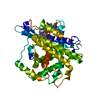
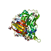
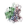
 Shewanella oneidensis (bacteria)
 X-RAY DIFFRACTION /
  Authors
 Citation
 Structure visualization
 Downloads & links
 Other downloads


 PDBj
PDBj
 Assembly


 Mass: 40.078 Da / Num. of mol.: 1 / Source method: obtained synthetically / Formula: Ca
 Mass: 22.990 Da / Num. of mol.: 3 / Source method: obtained synthetically / Formula: Na
 Mass: 59.044 Da / Num. of mol.: 6 / Source method: obtained synthetically / Formula: C2H3O2
 Mass: 92.094 Da / Num. of mol.: 1 / Source method: obtained synthetically / Formula: C3H8O3
 Sample preparation
 / Beamline: BL11-1 / Wavelength: 0.91837, 0.97931, 0.97885
 Processing