 Movie
Movie Controller

[English] 日本語
 Yorodumi
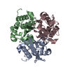
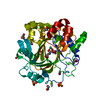
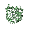
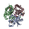
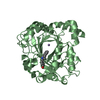
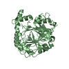
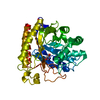
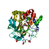
- PDB-2op8: Crystal Structure of YwhB- Homologue of 4-Oxalocrotonate Tautomerase -
+ Open data

- Basic information
| Entry | Database: PDB / ID: 2op8 | ||||||
|---|---|---|---|---|---|---|---|
| Title | Crystal Structure of YwhB- Homologue of 4-Oxalocrotonate Tautomerase | ||||||
 Components Components | Probable tautomerase ywhB | ||||||
 Keywords Keywords | ISOMERASE / 4-OT / tautomerase | ||||||
| Function / homology |  Function and homology information Function and homology information | ||||||
| Biological species |  | ||||||
| Method |  X-RAY DIFFRACTION / MOLECULAR REPLACEMENT / Resolution: 2.5 Å X-RAY DIFFRACTION / MOLECULAR REPLACEMENT / Resolution: 2.5 Å | ||||||
 Authors Authors | Hackert, M.L. / Whitman, C.P. / Almrud, J.J. | ||||||
 Citation Citation | Journal: TO BE PUBLISHED Title: The Crystal Structure of YwhB, a 4-Oxalocrotonate Tautomerase Homologue from Bacillus subtilis: The Structural Basis for Catalysis, Inhibition, and Reaction Stereoselectivity Authors: Hackert, M.L. / Whitman, C.P. / Almrud, J.J. / Dasgupta, R. / Wang, S.C. / Johnson, W.H. | ||||||
| History |
|
- Structure visualization
| Structure viewer | Molecule: MolmilJmol/JSmol |
|---|
- Downloads & links
-Download
| PDBx/mmCIF format | 2op8.cif.gz | 35.8 KB | Display | PDBx/mmCIF format |
|---|---|---|---|---|
| PDB format | pdb2op8.ent.gz | 25.5 KB | Display | PDB format |
| PDBx/mmJSON format | 2op8.json.gz | Tree view | PDBx/mmJSON format | |
| Others |  Other downloads Other downloads |
-Validation report
| Arichive directory | https://data.pdbj.org/pub/pdb/validation_reports/op/2op8ftp://data.pdbj.org/pub/pdb/validation_reports/op/2op8 | HTTPS FTP |
|---|
-Related structure data
| Related structure data |  1bjpS S: Starting model for refinement |
|---|---|
| Similar structure data |


-Links
 PDBj
PDBj- Assembly
| Deposited unit | 
| |||||||||
|---|---|---|---|---|---|---|---|---|---|---|
| 1 | 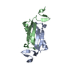
| |||||||||
| Unit cell |
| |||||||||
| Components on special symmetry positions |
|
-Components
| #1: Protein | Mass: 7026.026 Da / Num. of mol.: 2 Source method: isolated from a genetically manipulated source Source: (gene. exp.) References: UniProt: P70994, Isomerases; Intramolecular oxidoreductases; Interconverting keto- and enol-groups #2: Water | ChemComp-HOH / |  Mass: 18.015 Da / Num. of mol.: 48 / Source method: isolated from a natural source / Formula: H2O Mass: 18.015 Da / Num. of mol.: 48 / Source method: isolated from a natural source / Formula: H2O |
|---|
-Experimental details
-Experiment
| Experiment | Method: X-RAY DIFFRACTION / Number of used crystals: 1 |
|---|
- Sample preparation
| Crystal | Density Matthews: 4.23 Å3/Da / Density % sol: 70.9 % |
|---|---|
| Crystal grow | Temperature: 277 K / Method: vapor diffusion, sitting drop / pH: 7.3 Details: protein (25 mg/ml) buffered in 50 mM HEPES, pH 7.3, 5uL of protein solution mixed with 5uL of 50% methyl-pentanediol (MPD), 0.2M (NH4)H2PO4, pH 6.5, VAPOR DIFFUSION, SITTING DROP, temperature 277K, pH 7.30 |
-Data collection
| Diffraction | Mean temperature: 98 K |
|---|---|
| Diffraction source | Source: ROTATING ANODE / Type: RIGAKU RU200 |
| Detector | Type: RIGAKU RAXIS IV / Detector: IMAGE PLATE |
| Radiation | Protocol: SINGLE WAVELENGTH / Monochromatic (M) / Laue (L): M / Scattering type: x-ray |
| Radiation wavelength | Relative weight: 1 |
| Reflection | Resolution: 2.5→30 Å / Num. obs: 8746 / % possible obs: 99.9 % / Redundancy: 68.27 % / Biso Wilson estimate: 35 Å2 / Rsym value: 0.086 / Net I/σ(I): 21.5 |
| Reflection shell | Resolution: 2.5→2.59 Å / Mean I/σ(I) obs: 6 / Rsym value: 0.483 / % possible all: 0.9 |
- Processing
| Software |
| ||||||||||||||||||||||||||||||||||||||||||||||||||||||||||||
|---|---|---|---|---|---|---|---|---|---|---|---|---|---|---|---|---|---|---|---|---|---|---|---|---|---|---|---|---|---|---|---|---|---|---|---|---|---|---|---|---|---|---|---|---|---|---|---|---|---|---|---|---|---|---|---|---|---|---|---|---|---|
| Refinement | Method to determine structure: MOLECULAR REPLACEMENT Starting model: PDB ENTRY 1BJP Resolution: 2.5→30 Å / Isotropic thermal model: ISOTROPIC / Cross valid method: THROUGHOUT / σ(F): 2 / Stereochemistry target values: ENGH & HUBER
| ||||||||||||||||||||||||||||||||||||||||||||||||||||||||||||
| Displacement parameters | Biso mean: 41 Å2 | ||||||||||||||||||||||||||||||||||||||||||||||||||||||||||||
| Refine analyze |
| ||||||||||||||||||||||||||||||||||||||||||||||||||||||||||||
| Refinement step | Cycle: LAST / Resolution: 2.5→30 Å
| ||||||||||||||||||||||||||||||||||||||||||||||||||||||||||||
| Refine LS restraints |
| ||||||||||||||||||||||||||||||||||||||||||||||||||||||||||||
| LS refinement shell | Resolution: 2.5→2.61 Å / Rfactor Rfree error: 0.038
|
