 Movie
Movie Controller

[English] 日本語
 Yorodumi
- PDB-2ofw: Crystal structure of the APSK domain of human PAPSS1 complexed wi... -
+ Open data

- Basic information
| Entry | Database: PDB / ID: 2ofw | ||||||
|---|---|---|---|---|---|---|---|
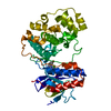
| Title | Crystal structure of the APSK domain of human PAPSS1 complexed with 2 APS molecules | ||||||
 Components Components | APS kinase domain of the PAPS synthetase 1 | ||||||
 Keywords Keywords | TRANSFERASE / nucleotide kinase | ||||||
| Function / homology |  Function and homology information Function and homology information3'-phosphoadenosine 5'-phosphosulfate biosynthetic process / Transport and metabolism of PAPS / sulfate adenylyltransferase / adenylyl-sulfate kinase / adenylylsulfate kinase activity / sulfate adenylyltransferase (ATP) activity / Metabolism of ingested H2SeO4 and H2SeO3 into H2Se / sulfate assimilation / nucleotidyltransferase activity / skeletal system development ...3'-phosphoadenosine 5'-phosphosulfate biosynthetic process / Transport and metabolism of PAPS / sulfate adenylyltransferase / adenylyl-sulfate kinase / adenylylsulfate kinase activity / sulfate adenylyltransferase (ATP) activity / Metabolism of ingested H2SeO4 and H2SeO3 into H2Se / sulfate assimilation / nucleotidyltransferase activity / skeletal system development / Signaling by BRAF and RAF1 fusions / protein homodimerization activity / ATP binding / nucleus / cytosol Similarity search - Function | ||||||
| Biological species |  Homo sapiens (human) Homo sapiens (human) | ||||||
| Method |  X-RAY DIFFRACTION / SYNCHROTRON / MOLECULAR REPLACEMENT / Resolution: 2.05 Å X-RAY DIFFRACTION / SYNCHROTRON / MOLECULAR REPLACEMENT / Resolution: 2.05 Å | ||||||
 Authors Authors | Sekulic, N. / Lavie, A. | ||||||
 Citation Citation | Journal: J.Mol.Biol. / Year: 2007 Title: Elucidation of the Active Conformation of the APS-Kinase Domain of Human PAPS Synthetase 1. Authors: Sekulic, N. / Dietrich, K. / Paarmann, I. / Ort, S. / Konrad, M. / Lavie, A. | ||||||
| History |
|
- Structure visualization
| Structure viewer | Molecule: MolmilJmol/JSmol |
|---|
- Downloads & links
-Download
| PDBx/mmCIF format | 2ofw.cif.gz | 346.2 KB | Display | PDBx/mmCIF format |
|---|---|---|---|---|
| PDB format | pdb2ofw.ent.gz | 279 KB | Display | PDB format |
| PDBx/mmJSON format | 2ofw.json.gz | Tree view | PDBx/mmJSON format | |
| Others |  Other downloads Other downloads |
-Validation report
| Arichive directory | https://data.pdbj.org/pub/pdb/validation_reports/of/2ofwftp://data.pdbj.org/pub/pdb/validation_reports/of/2ofw | HTTPS FTP |
|---|
-Related structure data
| Related structure data |  2ofxC 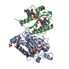 1m7gS S: Starting model for refinement C: citing same article ( |
|---|---|
| Similar structure data |


-Links
 PDBj
PDBj
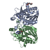
- Assembly
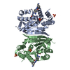
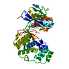
| Deposited unit | 
| ||||||||
|---|---|---|---|---|---|---|---|---|---|
| 1 | 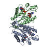
| ||||||||
| 2 | 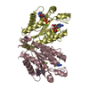
| ||||||||
| 3 | 
| ||||||||
| 4 | 
| ||||||||
| Unit cell |
|
-Components
| #1: Protein | Mass: 23100.082 Da / Num. of mol.: 8 / Fragment: APS kinse domain (Residues 1-227) Source method: isolated from a genetically manipulated source Source: (gene. exp.) Homo sapiens (human) / Gene: PAPSS1, ATPSK1, PAPSS / Plasmid: pGEX-RB with engineered TEV cutting site / Production host:  #2: Chemical | ChemComp-ADX /   Type: RNA linking / Mass: 427.284 Da / Num. of mol.: 16 / Source method: obtained synthetically / Formula: C10H14N5O10PS Type: RNA linking / Mass: 427.284 Da / Num. of mol.: 16 / Source method: obtained synthetically / Formula: C10H14N5O10PS#3: Chemical | ChemComp-MG /   Mass: 24.305 Da / Num. of mol.: 6 / Source method: obtained synthetically / Formula: Mg Mass: 24.305 Da / Num. of mol.: 6 / Source method: obtained synthetically / Formula: Mg#4: Water | ChemComp-HOH / |  Mass: 18.015 Da / Num. of mol.: 832 / Source method: isolated from a natural source / Formula: H2O Mass: 18.015 Da / Num. of mol.: 832 / Source method: isolated from a natural source / Formula: H2O |
|---|
-Experimental details
-Experiment
| Experiment | Method: X-RAY DIFFRACTION / Number of used crystals: 1 |
|---|
- Sample preparation
| Crystal | Density Matthews: 2.28 Å3/Da / Density % sol: 46 % |
|---|---|
| Crystal grow | Temperature: 295 K / Method: vapor diffusion, hanging drop Details: reservoir: 18-20% PEG 3350, 0.25 M calcium acetate protein solution: 5-8 mg/ml protein, 3 mM APS, 5 mM MgCl2, 50 mM Tris pH 7.5, 50 mM KCl, VAPOR DIFFUSION, HANGING DROP, temperature 295K |
-Data collection
| Diffraction | Mean temperature: 100 K |
|---|---|
| Diffraction source | Source: SYNCHROTRON / Site: APS  / Beamline: 22-BM / Wavelength: 0.9594 Å / Beamline: 22-BM / Wavelength: 0.9594 Å |
| Detector | Type: MARMOSAIC 225 mm CCD / Detector: CCD / Date: Mar 17, 2004 |
| Radiation | Protocol: SINGLE WAVELENGTH / Monochromatic (M) / Laue (L): M / Scattering type: x-ray |
| Radiation wavelength | Wavelength: 0.9594 Å / Relative weight: 1 |
| Reflection | Resolution: 2.05→10 Å / Num. all: 121588 / Num. obs: 101698 / % possible obs: 83.6 % / Observed criterion σ(F): 0 / Redundancy: 3.7 % / Rmerge(I) obs: 0.095 / Rsym value: 0.082 / Net I/σ(I): 12.4 |
| Reflection shell | Resolution: 2.05→2.1 Å / Redundancy: 2.5 % / Rmerge(I) obs: 0.301 / Mean I/σ(I) obs: 4.36 / Num. unique all: 7260 / Rsym value: 0.24 / % possible all: 92.8 |
-Phasing
| Phasing MR | Rfactor: 0.532 / Cor.coef. Fo:Fc: 0.272
|
|---|
- Processing
| Software |
| |||||||||||||||||||||||||||||||||||||||||||||||||||||||||||||||||||||||||||||||||||||||||||||||
|---|---|---|---|---|---|---|---|---|---|---|---|---|---|---|---|---|---|---|---|---|---|---|---|---|---|---|---|---|---|---|---|---|---|---|---|---|---|---|---|---|---|---|---|---|---|---|---|---|---|---|---|---|---|---|---|---|---|---|---|---|---|---|---|---|---|---|---|---|---|---|---|---|---|---|---|---|---|---|---|---|---|---|---|---|---|---|---|---|---|---|---|---|---|---|---|---|
| Refinement | Method to determine structure: MOLECULAR REPLACEMENT Starting model: pdb id 1M7G Resolution: 2.05→10 Å / Cor.coef. Fo:Fc: 0.925 / Cor.coef. Fo:Fc free: 0.877 / SU B: 6.244 / SU ML: 0.173 / Cross valid method: THROUGHOUT / σ(F): 0 / ESU R: 0.285 / ESU R Free: 0.234 / Stereochemistry target values: MAXIMUM LIKELIHOOD / Details: HYDROGENS HAVE BEEN ADDED IN THE RIDING POSITIONS
| |||||||||||||||||||||||||||||||||||||||||||||||||||||||||||||||||||||||||||||||||||||||||||||||
| Solvent computation | Ion probe radii: 0.8 Å / Shrinkage radii: 0.8 Å / VDW probe radii: 1.4 Å / Solvent model: MASK | |||||||||||||||||||||||||||||||||||||||||||||||||||||||||||||||||||||||||||||||||||||||||||||||
| Displacement parameters | Biso mean: 27.759 Å2
| |||||||||||||||||||||||||||||||||||||||||||||||||||||||||||||||||||||||||||||||||||||||||||||||
| Refinement step | Cycle: LAST / Resolution: 2.05→10 Å
| |||||||||||||||||||||||||||||||||||||||||||||||||||||||||||||||||||||||||||||||||||||||||||||||
| Refine LS restraints |
| |||||||||||||||||||||||||||||||||||||||||||||||||||||||||||||||||||||||||||||||||||||||||||||||
| LS refinement shell | Resolution: 2.05→2.101 Å / Total num. of bins used: 20
|
