SEQUENCE THE CONSTRUCT WAS EXPRESSED WITH A PURIFICATION TAG MGSDKIHHHHHHENLYFQG. THE TAG WAS ...SEQUENCE THE CONSTRUCT WAS EXPRESSED WITH A PURIFICATION TAG MGSDKIHHHHHHENLYFQG. THE TAG WAS REMOVED WITH TEV PROTEASE LEAVING ONLY A GLYCINE (0) FOLLOWED BY THE TARGET SEQUENCE. RESIDUE 56 IS MUTATED FROM LYSINE TO TYROSINE. RESIDUE 58 IS MUTATED FROM ASPARTIC ACID TO TYROSINE.
Resolution: 2.3→29.26 Å / Num. obs: 58378 / % possible obs: 93.5 % / Biso Wilson estimate: 40.054 Å2 / Rmerge(I) obs: 0.056 / Net I/σ(I): 9.71
Reflection shell
Diffraction-ID: 1
Resolution (Å)
Highest resolution (Å)
Rmerge(I) obs
Mean I/σ(I) obs
Num. measured obs
Num. unique all
% possible all
2.3-2.38
0.307
2.4
15585
9700
85.6
2.38-2.48
0.258
2.9
17936
11131
90.7
2.48-2.59
0.208
3.4
16853
10456
91.7
2.59-2.73
0.18
4.1
18030
11163
93.9
2.73-2.9
0.124
5.6
17756
10994
94.7
2.9-3.12
0.082
8.1
17743
10951
95.8
3.12-3.43
0.058
11.6
18016
11120
95.9
3.43-3.93
0.045
15.1
17886
11105
94.5
3.93
0.03
19.5
17773
11053
95.9
-
Phasing
Phasing
Method: MAD
-
Processing
Software
Name
Version
Classification
NB
MolProbity
3beta29
modelbuilding
SHELX
phasing
REFMAC
5.2.0005
refinement
XSCALE
datascaling
PDB_EXTRACT
2
dataextraction
XDS
datareduction
SHELXD
phasing
autoSHARP
phasing
Refinement
Method to determine structure: MAD / Resolution: 2.3→29.26 Å / Cor.coef. Fo:Fc: 0.956 / Cor.coef. Fo:Fc free: 0.938 / SU B: 13.344 / SU ML: 0.171 / TLS residual ADP flag: LIKELY RESIDUAL / Cross valid method: THROUGHOUT / σ(F): 0 / ESU R: 0.379 / ESU R Free: 0.221 Stereochemistry target values: MAXIMUM LIKELIHOOD WITH PHASES Details: 1.HYDROGENS HAVE BEEN ADDED IN THE RIDING POSITIONS. 2.ATOM RECORD CONTAINS RESIDUAL B FACTORS ONLY. 3.A MET-INHIBITION PROTOCOL WAS USED FOR SELENOMETHIONINE INCORPORATION DURING PROTEIN ...Details: 1.HYDROGENS HAVE BEEN ADDED IN THE RIDING POSITIONS. 2.ATOM RECORD CONTAINS RESIDUAL B FACTORS ONLY. 3.A MET-INHIBITION PROTOCOL WAS USED FOR SELENOMETHIONINE INCORPORATION DURING PROTEIN EXPRESSION. THE OCCUPANCY OF THE SE ATOMS IN THE MSE RESIDUES WAS REDUCED TO 0.75 TO ACCOUNT FOR THE REDUCED SCATTERING POWER DUE TO PARTIAL S-MET INCORPORATION. 4.PHOSPHATE ION IS MODELED AROUND RESIDUE 164,165 AND 166 IN EACH SUBUNIT. PEG 400 MOLECULES FROM CRYOPRECTANT ARE MODELED IN EACH SUBUNIT. 5.THERE IS UNKNOWN ELECTRON DENSITY NEAR TO RESIDUE 306 IN EACH SUBUNIT. 6.RESIDUE 84 IN SUBUNIT A, RESIDUE 83 IN SUBUNIT C AND D ARE DISORDERED AND NOT BUILT IN THIS MODEL.
Rfactor
Num. reflection
% reflection
Selection details
Rfree
0.214
2964
5.1 %
RANDOM
Rwork
0.175
-
-
-
obs
0.177
58346
98.73 %
-
Solvent computation
Ion probe radii: 0.8 Å / Shrinkage radii: 0.8 Å / VDW probe radii: 1.2 Å / Solvent model: MASK
Displacement parameters
Biso mean: 37.889 Å2
Baniso -1
Baniso -2
Baniso -3
1-
1.1 Å2
0 Å2
1.42 Å2
2-
-
1.11 Å2
0 Å2
3-
-
-
-1.57 Å2
Refinement step
Cycle: LAST / Resolution: 2.3→29.26 Å
Protein
Nucleic acid
Ligand
Solvent
Total
Num. atoms
10117
0
72
521
10710
Refine LS restraints
Refine-ID
Type
Dev ideal
Dev ideal target
Number
X-RAY DIFFRACTION
r_bond_refined_d
0.01
0.022
10380
X-RAY DIFFRACTION
r_bond_other_d
0.002
0.02
9811
X-RAY DIFFRACTION
r_angle_refined_deg
1.464
1.977
14169
X-RAY DIFFRACTION
r_angle_other_deg
0.89
3
22642
X-RAY DIFFRACTION
r_dihedral_angle_1_deg
3.226
5
1369
X-RAY DIFFRACTION
r_dihedral_angle_2_deg
32.992
23.867
375
X-RAY DIFFRACTION
r_dihedral_angle_3_deg
10.592
15
1576
X-RAY DIFFRACTION
r_dihedral_angle_4_deg
14.049
15
63
X-RAY DIFFRACTION
r_chiral_restr
0.079
0.2
1688
X-RAY DIFFRACTION
r_gen_planes_refined
0.004
0.02
11610
X-RAY DIFFRACTION
r_gen_planes_other
0.001
0.02
1915
X-RAY DIFFRACTION
r_nbd_refined
0.192
0.3
1976
X-RAY DIFFRACTION
r_nbd_other
0.154
0.3
9720
X-RAY DIFFRACTION
r_nbtor_refined
0.165
0.5
5090
X-RAY DIFFRACTION
r_nbtor_other
0.083
0.5
6013
X-RAY DIFFRACTION
r_xyhbond_nbd_refined
0.187
0.5
810
X-RAY DIFFRACTION
r_xyhbond_nbd_other
0.02
0.5
1
X-RAY DIFFRACTION
r_symmetry_vdw_refined
0.147
0.3
18
X-RAY DIFFRACTION
r_symmetry_vdw_other
0.151
0.3
84
X-RAY DIFFRACTION
r_symmetry_hbond_refined
0.168
0.5
12
X-RAY DIFFRACTION
r_mcbond_it
1.05
3
6998
X-RAY DIFFRACTION
r_mcbond_other
0.313
3
2821
X-RAY DIFFRACTION
r_mcangle_it
1.528
5
10934
X-RAY DIFFRACTION
r_scbond_it
2.758
8
3809
X-RAY DIFFRACTION
r_scangle_it
4.042
11
3235
Refine LS restraints NCS
Refine-ID: X-RAY DIFFRACTION
Ens-ID
Dom-ID
Auth asym-ID
Number
Type
Rms dev position (Å)
Weight position
1
1
A
1777
MEDIUMPOSITIONAL
0.37
0.5
1
2
B
1777
MEDIUMPOSITIONAL
0.41
0.5
1
3
C
1777
MEDIUMPOSITIONAL
0.4
0.5
1
4
D
1777
MEDIUMPOSITIONAL
0.43
0.5
1
1
A
1777
MEDIUMTHERMAL
0.38
2
1
2
B
1777
MEDIUMTHERMAL
0.43
2
1
3
C
1777
MEDIUMTHERMAL
0.37
2
1
4
D
1777
MEDIUMTHERMAL
0.39
2
2
1
A
3060
MEDIUMPOSITIONAL
0.37
0.5
2
2
B
3060
MEDIUMPOSITIONAL
0.39
0.5
2
3
C
3060
MEDIUMPOSITIONAL
0.32
0.5
2
4
D
3060
MEDIUMPOSITIONAL
0.36
0.5
2
1
A
3060
MEDIUMTHERMAL
0.57
2
2
2
B
3060
MEDIUMTHERMAL
0.58
2
2
3
C
3060
MEDIUMTHERMAL
0.54
2
2
4
D
3060
MEDIUMTHERMAL
0.58
2
LS refinement shell
Resolution: 2.3→2.36 Å / Total num. of bins used: 20
Rfactor
Num. reflection
% reflection
Rfree
0.258
193
-
Rwork
0.217
3965
-
obs
-
4158
95.19 %
Refinement TLS params.
Method: refined / Refine-ID: X-RAY DIFFRACTION
ID
L11 (°2)
L12 (°2)
L13 (°2)
L22 (°2)
L23 (°2)
L33 (°2)
S11 (Å °)
S12 (Å °)
S13 (Å °)
S21 (Å °)
S22 (Å °)
S23 (Å °)
S31 (Å °)
S32 (Å °)
S33 (Å °)
T11 (Å2)
T12 (Å2)
T13 (Å2)
T22 (Å2)
T23 (Å2)
T33 (Å2)
Origin x (Å)
Origin y (Å)
Origin z (Å)
1
2.3401
-0.7318
-1.064
3.1667
-0.084
5.3734
0.1557
-0.3307
0.3965
0.0777
-0.0481
-0.363
-0.4564
0.6603
-0.1076
0.056
-0.0846
0.0225
0.0281
-0.0724
-0.0727
-4.67
53.7296
43.0082
2
1.7256
-0.2577
-1.2358
1.853
0.1654
2.4865
0.2706
-0.0128
0.3178
0.2714
0.0405
0.1437
-0.5324
0.0493
-0.3112
-0.0358
-0.007
0.0913
-0.2561
-0.0194
-0.0956
-13.6294
59.4701
17.2379
3
1.4518
-0.1903
-0.711
4.7322
1.3552
5.9504
-0.0585
-0.0409
-0.3594
0.3383
0.1631
0.1083
0.3799
0.0008
-0.1046
-0.1152
0.0613
-0.0114
-0.1797
-0.0176
0.0234
-1.6981
16.7169
12.5788
4
1.8482
0.1535
-0.5459
1.2258
0.5398
1.8202
-0.0724
0.1673
-0.0753
0.0237
0.1285
-0.0714
-0.0777
0.2158
-0.0561
-0.2355
-0.0296
-0.0205
-0.1396
-0.062
-0.1918
2.8862
41.8262
1.264
5
1.4419
0.7623
0.2125
5.1712
1.3688
6.4247
0.0915
0.3458
-0.2964
-0.1067
-0.1484
0.273
0.609
-0.4181
0.0568
0.1051
-0.1086
0.0394
0.0371
-0.0663
0.0663
-44.5723
2.7549
22.2269
6
2.5853
-0.0184
-0.3157
1.0843
0.5147
4.2
0.0111
0.2391
0.3345
-0.2722
0.0116
-0.0436
-0.3519
-0.0878
-0.0227
-0.1558
-0.0327
-0.026
-0.1695
0.0142
-0.0953
-38.7318
28.3514
31.9444
7
2.1773
-0.1971
-0.3801
5.0429
2.9237
6.2192
-0.0241
-0.4518
-0.2279
0.2823
0.4533
-0.2562
0.3698
0.9904
-0.4293
0.0494
-0.0354
-0.0234
0.1919
-0.0379
-0.0622
-46.1479
5.2727
69.8458
8
1.4273
-0.6328
-1.7826
1.2187
1.2444
4.0561
0.0252
0.0737
0.1912
-0.0068
-0.1119
0.005
-0.0054
-0.6527
0.0867
-0.2767
-0.027
-0.0516
-0.0111
-0.083
-0.121
-53.6888
28.8092
56.8713
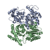
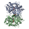
Refinement TLS group
Refine-ID: X-RAY DIFFRACTION / Selection: ALL
ID
Refine TLS-ID
Auth asym-ID
Label asym-ID
Auth seq-ID
Label seq-ID
1
1
A
A
6 - 134
7 - 135
2
2
A
A
135 - 348
136 - 349
3
3
B
B
6 - 134
7 - 135
4
4
B
B
135 - 348
136 - 349
5
5
C
C
3 - 134
4 - 135
6
6
C
C
135 - 348
136 - 349
7
7
D
D
2 - 134
3 - 135
8
8
D
D
135 - 348
136 - 349
+
About Yorodumi
-
News
-
Feb 9, 2022. New format data for meta-information of EMDB entries
New format data for meta-information of EMDB entries
Version 3 of the EMDB header file is now the official format.
The previous official version 1.9 will be removed from the archive.
In the structure databanks used in Yorodumi, some data are registered as the other names, "COVID-19 virus" and "2019-nCoV". Here are the details of the virus and the list of structure data.
Jan 31, 2019. EMDB accession codes are about to change! (news from PDBe EMDB page)
EMDB accession codes are about to change! (news from PDBe EMDB page)
The allocation of 4 digits for EMDB accession codes will soon come to an end. Whilst these codes will remain in use, new EMDB accession codes will include an additional digit and will expand incrementally as the available range of codes is exhausted. The current 4-digit format prefixed with “EMD-” (i.e. EMD-XXXX) will advance to a 5-digit format (i.e. EMD-XXXXX), and so on. It is currently estimated that the 4-digit codes will be depleted around Spring 2019, at which point the 5-digit format will come into force.
The EM Navigator/Yorodumi systems omit the EMD- prefix.
Related info.:Q: What is EMD? / ID/Accession-code notation in Yorodumi/EM Navigator
Yorodumi is a browser for structure data from EMDB, PDB, SASBDB, etc.
This page is also the successor to EM Navigator detail page, and also detail information page/front-end page for Omokage search.
The word "yorodu" (or yorozu) is an old Japanese word meaning "ten thousand". "mi" (miru) is to see.
Related info.:EMDB / PDB / SASBDB / Comparison of 3 databanks / Yorodumi Search / Aug 31, 2016. New EM Navigator & Yorodumi / Yorodumi Papers / Jmol/JSmol / Function and homology information / Changes in new EM Navigator and Yorodumi
 Movie
Movie Controller

 Yorodumi
 Open data
 Basic information
 Components
 Keywords
 Function and homology information
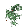
 Anabaena variabilis (bacteria)
 X-RAY DIFFRACTION /
  Authors
 Citation
 Structure visualization
 Downloads & links
 Other downloads


 PDBj
PDBj Assembly


 Mass: 94.971 Da / Num. of mol.: 4 / Source method: obtained synthetically / Formula: PO4
 Mass: 194.226 Da / Num. of mol.: 4 / Source method: obtained synthetically / Formula: C8H18O5 / Comment: precipitant*YM
 Mass: 18.015 Da / Num. of mol.: 521 / Source method: isolated from a natural source / Formula: H2O
 Sample preparation
 / Beamline: 5.0.3 / Wavelength: 1.0000, 0.9806, 0.9805
 Processing