| 登録情報 | データベース: PDB / ID: 2o7p
|
|---|
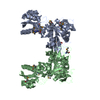
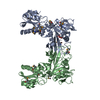
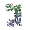
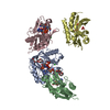
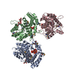
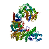
| タイトル | The crystal structure of RibD from Escherichia coli in complex with the oxidised NADP+ cofactor in the active site of the reductase domain |
|---|
 要素 要素 | Riboflavin biosynthesis protein ribD |
|---|
 キーワード キーワード | HYDROLASE / OXIDOREDUCTASE / NADP+ complex / alpha and beta class with mainly parallell beta strands / Structural Genomics / HTP-protein Escherichia coli / Structural Proteomics in Europe / SPINE |
|---|
| 機能・相同性 |  機能・相同性情報 機能・相同性情報
5-amino-6-(5-phosphoribosylamino)uracil reductase / diaminohydroxyphosphoribosylaminopyrimidine deaminase / diaminohydroxyphosphoribosylaminopyrimidine deaminase activity / 5-amino-6-(5-phosphoribosylamino)uracil reductase activity / riboflavin biosynthetic process / NADP binding / zinc ion binding類似検索 - 分子機能 Riboflavin biosynthesis protein RibD / Riboflavin-specific deaminase, C-terminal / : / Bacterial bifunctional deaminase-reductase, C-terminal / RibD C-terminal domain / Cytidine and deoxycytidylate deaminase zinc-binding region / Cytidine Deaminase, domain 2 / Cytidine Deaminase; domain 2 / APOBEC/CMP deaminase, zinc-binding / Cytidine and deoxycytidylate deaminases zinc-binding region signature. ...Riboflavin biosynthesis protein RibD / Riboflavin-specific deaminase, C-terminal / : / Bacterial bifunctional deaminase-reductase, C-terminal / RibD C-terminal domain / Cytidine and deoxycytidylate deaminase zinc-binding region / Cytidine Deaminase, domain 2 / Cytidine Deaminase; domain 2 / APOBEC/CMP deaminase, zinc-binding / Cytidine and deoxycytidylate deaminases zinc-binding region signature. / Cytidine and deoxycytidylate deaminase domain / Cytidine and deoxycytidylate deaminases domain profile. / Cytidine deaminase-like / Dihydrofolate Reductase, subunit A / Dihydrofolate Reductase, subunit A / Dihydrofolate reductase-like domain superfamily / 3-Layer(aba) Sandwich / Alpha Beta類似検索 - ドメイン・相同性 NADP NICOTINAMIDE-ADENINE-DINUCLEOTIDE PHOSPHATE / Riboflavin biosynthesis protein RibD / Riboflavin biosynthesis protein RibD類似検索 - 構成要素 |
|---|
| 生物種 |   Escherichia coli (大腸菌) Escherichia coli (大腸菌) |
|---|
| 手法 |  X線回折 / シンクロトロン / 分子置換 / 解像度: 3 Å X線回折 / シンクロトロン / 分子置換 / 解像度: 3 Å |
|---|
 データ登録者 データ登録者 | Moche, M. / Stenmark, P. / Gurmu, D. / Nordlund, P. / Structural Proteomics in Europe (SPINE) |
|---|
 引用 引用 | ジャーナル: J.Mol.Biol. / 年: 2007
タイトル: The crystal structure of the bifunctional deaminase/reductase RibD of the riboflavin biosynthetic pathway in Escherichia coli: implications for the reductive mechanism.
著者: Stenmark, P. / Moche, M. / Gurmu, D. / Nordlund, P. |
|---|
| 履歴 | | 登録 | 2006年12月11日 | 登録サイト: RCSB / 処理サイト: PDBJ |
|---|
| 改定 1.0 | 2007年2月13日 | Provider: repository / タイプ: Initial release |
|---|
| 改定 1.1 | 2008年5月1日 | Group: Version format compliance |
|---|
| 改定 1.2 | 2011年7月13日 | Group: Source and taxonomy / Version format compliance |
|---|
| 改定 1.3 | 2015年6月3日 | Group: Database references |
|---|
| 改定 1.4 | 2023年10月25日 | Group: Data collection / Database references ...Data collection / Database references / Derived calculations / Refinement description
カテゴリ: chem_comp_atom / chem_comp_bond ...chem_comp_atom / chem_comp_bond / database_2 / pdbx_initial_refinement_model / struct_conn / struct_ref_seq_dif / struct_site
Item: _database_2.pdbx_DOI / _database_2.pdbx_database_accession ..._database_2.pdbx_DOI / _database_2.pdbx_database_accession / _struct_conn.pdbx_leaving_atom_flag / _struct_ref_seq_dif.details / _struct_site.pdbx_auth_asym_id / _struct_site.pdbx_auth_comp_id / _struct_site.pdbx_auth_seq_id |
|---|
| 改定 1.5 | 2023年11月15日 | Group: Data collection / カテゴリ: chem_comp_atom / chem_comp_bond / Item: _chem_comp_atom.atom_id / _chem_comp_bond.atom_id_2 |
|---|
| 改定 1.6 | 2024年10月30日 | Group: Structure summary
カテゴリ: pdbx_entry_details / pdbx_modification_feature |
|---|
|
|---|
 ムービー
ムービー コントローラー


 データを開く
 基本情報
基本情報 構造の表示
構造の表示 ダウンロードとリンク
 その他のダウンロード


 PDBj
PDBj
 集合体
集合体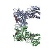

 分子量: 743.405 Da / 分子数: 2 / 由来タイプ: 合成 / 式: C21H28N7O17P3
 試料調製
試料調製 / ビームライン: ID29 / 波長: 1.2836 Å
 解析
解析