 Movie
Movie Controller


+ Open data

- Basic information
| Entry | Database: PDB / ID: 2o6f | ||||||
|---|---|---|---|---|---|---|---|
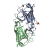
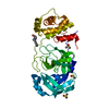
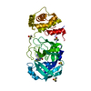
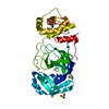
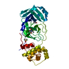
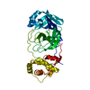
| Title | Structure of metal- free rTp34 from Treponema pallidum | ||||||
 Components Components | 34 kDa membrane antigen | ||||||
 Keywords Keywords | MEMBRANE PROTEIN / PROTEIN BINDING / Ig-fold / syphilis / metal-ion binding / dimer | ||||||
| Function / homology |  Function and homology information Function and homology information | ||||||
| Biological species |  Treponema pallidum (bacteria) Treponema pallidum (bacteria) | ||||||
| Method |  X-RAY DIFFRACTION / SYNCHROTRON / FOURIER SYNTHESIS / Resolution: 1.63 Å X-RAY DIFFRACTION / SYNCHROTRON / FOURIER SYNTHESIS / Resolution: 1.63 Å | ||||||
 Authors Authors | Machius, M. / Brautigam, C.A. / Deka, R.K. / Tomchick, D.R. / Lumpkins, S.B. / Norgard, M.V. | ||||||
 Citation Citation | Journal: J.Biol.Chem. / Year: 2007 Title: Crystal structure of the Tp34 (TP0971) lipoprotein of treponema pallidum: implications of its metal-bound state and affinity for human lactoferrin. Authors: Deka, R.K. / Brautigam, C.A. / Tomson, F.L. / Lumpkins, S.B. / Tomchick, D.R. / Machius, M. / Norgard, M.V. | ||||||
| History |
|
- Structure visualization
| Structure viewer | Molecule: MolmilJmol/JSmol |
|---|
- Downloads & links
-Download
| PDBx/mmCIF format | 2o6f.cif.gz | 83.6 KB | Display | PDBx/mmCIF format |
|---|---|---|---|---|
| PDB format | pdb2o6f.ent.gz | 63 KB | Display | PDB format |
| PDBx/mmJSON format | 2o6f.json.gz | Tree view | PDBx/mmJSON format | |
| Others |  Other downloads Other downloads |
-Validation report
| Summary document | 2o6f_validation.pdf.gz | 462.3 KB | Display | wwPDB validaton report |
|---|---|---|---|---|
| Full document | 2o6f_full_validation.pdf.gz | 465.2 KB | Display | |
| Data in XML | 2o6f_validation.xml.gz | 16.7 KB | Display | |
| Data in CIF | 2o6f_validation.cif.gz | 24.1 KB | Display | |
| Arichive directory | https://data.pdbj.org/pub/pdb/validation_reports/o6/2o6fftp://data.pdbj.org/pub/pdb/validation_reports/o6/2o6f | HTTPS FTP |
-Related structure data


-Links
 PDBj
PDBj


- Assembly
| Deposited unit | 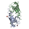
| ||||||||
|---|---|---|---|---|---|---|---|---|---|
| 1 |
| ||||||||
| Unit cell |
|
-Components
| #1: Protein | Mass: 20443.449 Da / Num. of mol.: 2 / Mutation: V16G, F17A, S18M, A19G, C20S Source method: isolated from a genetically manipulated source Source: (gene. exp.) Treponema pallidum (bacteria) / Gene: tpd / Plasmid: pProExHTa / Production host: #2: Chemical |   Mass: 96.063 Da / Num. of mol.: 2 / Source method: obtained synthetically / Formula: SO4 Mass: 96.063 Da / Num. of mol.: 2 / Source method: obtained synthetically / Formula: SO4#3: Chemical |   Mass: 35.453 Da / Num. of mol.: 3 / Source method: obtained synthetically / Formula: Cl Mass: 35.453 Da / Num. of mol.: 3 / Source method: obtained synthetically / Formula: Cl#4: Chemical |   Mass: 62.068 Da / Num. of mol.: 2 / Source method: obtained synthetically / Formula: C2H6O2 Mass: 62.068 Da / Num. of mol.: 2 / Source method: obtained synthetically / Formula: C2H6O2#5: Water | ChemComp-HOH / |  Mass: 18.015 Da / Num. of mol.: 240 / Source method: isolated from a natural source / Formula: H2O Mass: 18.015 Da / Num. of mol.: 240 / Source method: isolated from a natural source / Formula: H2O |
|---|
-Experimental details
-Experiment
| Experiment | Method: X-RAY DIFFRACTION / Number of used crystals: 1 |
|---|
- Sample preparation
| Crystal | Density Matthews: 2.09 Å3/Da / Density % sol: 41.06 % |
|---|---|
| Crystal grow | Temperature: 293 K / Method: vapor diffusion, hanging drop / pH: 9 Details: 2.4 M Ammonium sulfate, 0.1 M Bicine, pH 9.0, VAPOR DIFFUSION, HANGING DROP, temperature 293K |
-Data collection
| Diffraction | Mean temperature: 100 K |
|---|---|
| Diffraction source | Source: SYNCHROTRON / Site: APS  / Beamline: 19-BM / Wavelength: 0.97899 Å / Beamline: 19-BM / Wavelength: 0.97899 Å |
| Detector | Type: CUSTOM-MADE / Detector: CCD / Date: Jul 14, 2006 |
| Radiation | Monochromator: Double-crystal Si 111 / Protocol: SINGLE WAVELENGTH / Monochromatic (M) / Laue (L): M / Scattering type: x-ray |
| Radiation wavelength | Wavelength: 0.97899 Å / Relative weight: 1 |
| Reflection | Resolution: 1.63→26.03 Å / Num. all: 41967 / Num. obs: 41967 / % possible obs: 95.3 % / Observed criterion σ(I): -3 / Redundancy: 4.1 % / Biso Wilson estimate: 25 Å2 / Rsym value: 0.038 / Net I/σ(I): 32 |
| Reflection shell | Resolution: 1.63→1.66 Å / Redundancy: 3.7 % / Mean I/σ(I) obs: 2.2 / Num. unique all: 2016 / Rsym value: 0.487 / % possible all: 94.1 |
- Processing
| Software |
| ||||||||||||||||||||||||||||||||||||||||||||||||||||||||||||||||||||||||||||||||||||||||||
|---|---|---|---|---|---|---|---|---|---|---|---|---|---|---|---|---|---|---|---|---|---|---|---|---|---|---|---|---|---|---|---|---|---|---|---|---|---|---|---|---|---|---|---|---|---|---|---|---|---|---|---|---|---|---|---|---|---|---|---|---|---|---|---|---|---|---|---|---|---|---|---|---|---|---|---|---|---|---|---|---|---|---|---|---|---|---|---|---|---|---|---|
| Refinement | Method to determine structure: FOURIER SYNTHESIS Starting model: native protein Resolution: 1.63→26.03 Å / Cor.coef. Fo:Fc: 0.964 / Cor.coef. Fo:Fc free: 0.955 / SU B: 3.8 / SU ML: 0.067 / TLS residual ADP flag: LIKELY RESIDUAL / Cross valid method: THROUGHOUT / σ(F): 0 / ESU R: 0.099 / ESU R Free: 0.097 / Stereochemistry target values: MAXIMUM LIKELIHOOD / Details: HYDROGENS HAVE BEEN ADDED IN THE RIDING POSITIONS
| ||||||||||||||||||||||||||||||||||||||||||||||||||||||||||||||||||||||||||||||||||||||||||
| Solvent computation | Ion probe radii: 0.8 Å / Shrinkage radii: 0.8 Å / VDW probe radii: 1.4 Å / Solvent model: MASK | ||||||||||||||||||||||||||||||||||||||||||||||||||||||||||||||||||||||||||||||||||||||||||
| Displacement parameters | Biso mean: 25.686 Å2
| ||||||||||||||||||||||||||||||||||||||||||||||||||||||||||||||||||||||||||||||||||||||||||
| Refine analyze | Luzzati coordinate error obs: 0.231 Å | ||||||||||||||||||||||||||||||||||||||||||||||||||||||||||||||||||||||||||||||||||||||||||
| Refinement step | Cycle: LAST / Resolution: 1.63→26.03 Å
| ||||||||||||||||||||||||||||||||||||||||||||||||||||||||||||||||||||||||||||||||||||||||||
| Refine LS restraints |
| ||||||||||||||||||||||||||||||||||||||||||||||||||||||||||||||||||||||||||||||||||||||||||
| LS refinement shell | Resolution: 1.63→1.669 Å / Total num. of bins used: 20
| ||||||||||||||||||||||||||||||||||||||||||||||||||||||||||||||||||||||||||||||||||||||||||
| Refinement TLS params. | Method: refined / Refine-ID: X-RAY DIFFRACTION
| ||||||||||||||||||||||||||||||||||||||||||||||||||||||||||||||||||||||||||||||||||||||||||
| Refinement TLS group | Refine-ID: X-RAY DIFFRACTION / Selection: ALL / Auth seq-ID: 29 - 185 / Label seq-ID: 33 - 189
|
