 Movie
Movie Controller

[English] 日本語
 Yorodumi
- PDB-2o4f: Structure of a parallel-stranded guanine tetraplex crystallised w... -
+ Open data

- Basic information
| Entry | Database: PDB / ID: 2o4f | ||||||||||||||||||
|---|---|---|---|---|---|---|---|---|---|---|---|---|---|---|---|---|---|---|---|
| Title | Structure of a parallel-stranded guanine tetraplex crystallised with monovalent ions | ||||||||||||||||||
 Components Components | 5'-D(* Keywords KeywordsDNA / QUADRUPLE HELIX / PARALLEL-STRANDED | Function / homology | : / DNA |  Function and homology information Function and homology informationMethod |  X-RAY DIFFRACTION / SYNCHROTRON / MOLECULAR REPLACEMENT / Resolution: 1.5 Å X-RAY DIFFRACTION / SYNCHROTRON / MOLECULAR REPLACEMENT / Resolution: 1.5 Å  Authors AuthorsCreze, C. / Rinaldi, B. / Haser, R. / Bouvet, P. / Gouet, P. |  CitationJournal: Acta Crystallogr.,Sect.D / Year: 2007 CitationJournal: Acta Crystallogr.,Sect.D / Year: 2007Title: Structure of a d(TGGGGT) quadruplex crystallized in the presence of Li+ ions. Authors: Creze, C. / Rinaldi, B. / Haser, R. / Bouvet, P. / Gouet, P. History |
|
- Structure visualization
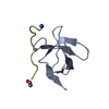
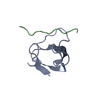
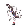
| Structure viewer | Molecule: MolmilJmol/JSmol |
|---|
- Downloads & links
-Download
| PDBx/mmCIF format | 2o4f.cif.gz | 40.7 KB | Display | PDBx/mmCIF format |
|---|---|---|---|---|
| PDB format | pdb2o4f.ent.gz | 29.4 KB | Display | PDB format |
| PDBx/mmJSON format | 2o4f.json.gz | Tree view | PDBx/mmJSON format | |
| Others |  Other downloads Other downloads |
-Validation report
| Arichive directory | https://data.pdbj.org/pub/pdb/validation_reports/o4/2o4fftp://data.pdbj.org/pub/pdb/validation_reports/o4/2o4f | HTTPS FTP |
|---|
-Related structure data
| Related structure data |  352dS S: Starting model for refinement |
|---|---|
| Similar structure data |


-Links
 PDBj
PDBj


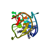
- Assembly
| Deposited unit | 
| ||||||||
|---|---|---|---|---|---|---|---|---|---|
| 1 | 
| ||||||||
| 2 | 
| ||||||||
| Unit cell |
|
-Components
| #1: DNA chain | Mass: 1880.251 Da / Num. of mol.: 8 / Source method: obtained synthetically #2: Chemical | ChemComp-NA /   Mass: 22.990 Da / Num. of mol.: 7 / Source method: obtained synthetically / Formula: Na Mass: 22.990 Da / Num. of mol.: 7 / Source method: obtained synthetically / Formula: Na#3: Chemical | ChemComp-LI / |   Mass: 6.941 Da / Num. of mol.: 1 / Source method: obtained synthetically / Formula: Li Mass: 6.941 Da / Num. of mol.: 1 / Source method: obtained synthetically / Formula: Li#4: Water | ChemComp-HOH / |  Mass: 18.015 Da / Num. of mol.: 228 / Source method: isolated from a natural source / Formula: H2O Mass: 18.015 Da / Num. of mol.: 228 / Source method: isolated from a natural source / Formula: H2O |
|---|
-Experimental details
-Experiment
| Experiment | Method: X-RAY DIFFRACTION / Number of used crystals: 1 |
|---|
- Sample preparation
| Crystal | Density Matthews: 1.82 Å3/Da / Density % sol: 32.49 % | ||||||||||||||||||||||||||||||||||||
|---|---|---|---|---|---|---|---|---|---|---|---|---|---|---|---|---|---|---|---|---|---|---|---|---|---|---|---|---|---|---|---|---|---|---|---|---|---|
| Crystal grow | Temperature: 277 K / Method: vapor diffusion, hanging drop / pH: 6 Details: 1.8 M lithium sulphate, 0.01 M magnesium sulphate, 0.05 M sodium cacodylate, pH 6.0, VAPOR DIFFUSION, HANGING DROP, temperature 277K | ||||||||||||||||||||||||||||||||||||
| Components of the solutions |
|
-Data collection
| Diffraction | Mean temperature: 100 K |
|---|---|
| Diffraction source | Source: SYNCHROTRON / Site: ESRF  / Beamline: ID23-2 / Wavelength: 0.8726 Å / Beamline: ID23-2 / Wavelength: 0.8726 Å |
| Detector | Type: MARRESEARCH / Detector: CCD / Date: Jul 16, 2006 / Details: mirrors |
| Radiation | Monochromator: horizontally diffracting Si (111) / Protocol: SINGLE WAVELENGTH / Monochromatic (M) / Laue (L): M / Scattering type: x-ray |
| Radiation wavelength | Wavelength: 0.8726 Å / Relative weight: 1 |
| Reflection | Resolution: 1.5→15 Å / Num. all: 18195 / Num. obs: 17547 / % possible obs: 96.4 % / Observed criterion σ(F): 0 / Observed criterion σ(I): -3 / Redundancy: 1.7 % / Biso Wilson estimate: 12 Å2 / Rsym value: 0.142 / Net I/σ(I): 7.05 |
| Reflection shell | Resolution: 1.5→1.59 Å / Redundancy: 1.7 % / Mean I/σ(I) obs: 2.71 / Num. unique all: 2977 / Rsym value: 0.227 / % possible all: 93.4 |
- Processing
| Software |
| ||||||||||||||||||||||||||||||||||||
|---|---|---|---|---|---|---|---|---|---|---|---|---|---|---|---|---|---|---|---|---|---|---|---|---|---|---|---|---|---|---|---|---|---|---|---|---|---|
| Refinement | Method to determine structure: MOLECULAR REPLACEMENT Starting model: PDB ENTRY 352D Resolution: 1.5→14.68 Å / Rfactor Rfree error: 0.009 / Data cutoff high absF: 690707.25 / Data cutoff low absF: 0 / Isotropic thermal model: RESTRAINED / Cross valid method: THROUGHOUT / σ(F): 0 / σ(I): -3 / Stereochemistry target values: Engh & Huber
| ||||||||||||||||||||||||||||||||||||
| Solvent computation | Solvent model: FLAT MODEL / Bsol: 37.627 Å2 / ksol: 0.355 e/Å3 | ||||||||||||||||||||||||||||||||||||
| Displacement parameters | Biso mean: 13 Å2
| ||||||||||||||||||||||||||||||||||||
| Refine analyze |
| ||||||||||||||||||||||||||||||||||||
| Refinement step | Cycle: LAST / Resolution: 1.5→14.68 Å
| ||||||||||||||||||||||||||||||||||||
| Refine LS restraints |
| ||||||||||||||||||||||||||||||||||||
| LS refinement shell | Resolution: 1.5→1.59 Å / Rfactor Rfree error: 0.028 / Total num. of bins used: 6
| ||||||||||||||||||||||||||||||||||||
| Xplor file |
|
